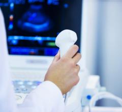
Schematic diagram of training and test flow. Image courtesy of Korea Institute of Science and Technology (KIST)
November 11, 2021 — Transcranial focused ultrasound can be used to treat degenerative movement disorders, intractable pain, and mental disorders by delivering ultrasound energy to a specific area of the brain without opening the skull. This treatment must be performed with an image-based technology that can locate the brain lesions. Doctors typically use computed tomography (CT) to obtain information about a patient's skull that is difficult to identify with magnetic resonance imaging (MRI) alone and to accurately focus the ultrasound on the lesions through the skull. However, there have been concerns about the safety of CT scans, during which radiation exposure is inevitable, especially in pediatric and pregnant patients.
Dr. Hyungmin Kim's team at the Bionics Research Center at the Korea Institute of Science and Technology (KIST, President Seok-Jin Yoon) developed an artificial intelligence technology to generate CT images based on MRI images and conducted a simulation experiment. The results showed that the transcranial focused ultrasound procedure could be performed with MRI alone.
Efforts have been made to obtain cranial information from MRI images, but special coils for the MRI or imaging protocols that are not widely available in the medical field are required. As an alternative, interest for acquiring artificial intelligence-based CT images has been high worldwide, but their clinical efficacy has not been proven. The KIST research team proved that CT images obtained by artificial intelligence have clinical utility.
The KIST research team developed a three-dimensional conditional adversarial generative network that learns the nonlinear CT transformation process from T1-weighted MRI images, which is one of the most commonly used images in the medical field. The team devised a loss function that minimizes the Hounsfield unit pixel variation error of the CT images, and also optimized the neural network performance by comparing the changes in quality of the synthetic CT images according to the normalization methods of MRI image signals, such as Z-score normalization and partial linear histogram matching normalization.
For safe and effective ultrasound treatment, it is imperative to understand each patient's skull density ratio and skull thickness in advance, and when these skull factors were obtained via the synthetic CT, both factors showed >0.90 correlation with the actual CT. There was no statistically significant difference. Moreover, when simulated ultrasound treatment was performed, the ultrasound focal distance had an error of less than 1 mm, the intracranial peak acoustic pressure had an error of approximately 3.1%, and the focal volume similarity was approximately 83%. This demonstrated that the transcranial focused ultrasound treatment system can be performed with only the MRI image.
Dr. Hyungmin Kim of KIST stated that “patients can receive focused ultrasound treatment without being worried about radiation exposure, and as the additional imaging and alignment processes can be omitted, this will reduce the staff's workload, leading to a reduction in time and economic costs.”He also stated that “through follow-up studies on identifying the error associated with the ultrasound parameters and transducers and understanding the possibility of artificial intelligence CT application in various parts of the body, we plan to continue developing the technology for its applicability in various treatment technologies.”
For more information: https://eng.kist.re.kr/kist_eng/main/

 April 25, 2024