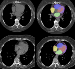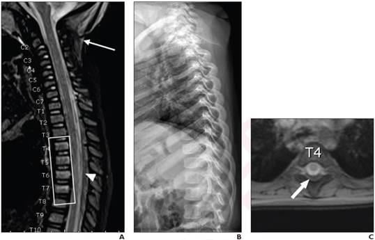
16-Month-Old Boy, Born at Gestational Age of 28 Weeks, Presenting With Unexplained Facial Burns: He also had unexplained rib, humerus, and tibial fractures, three skull fractures, severe acute hypoxic brain injury with transtentorial herniation, and intracranial subdural hematoma in interhemispheric falx and tentorium. Child diagnosed with abusive head trauma. A. Sagittal STIR image from whole-spine MRI demonstrates edema in nuchal ligament consistent with ligamentous injury (arrow) and epidural hematoma extending along thoracic spine (arrowhead), associated with compression fractures from T4 to T7 (rectangle). Epidural hematoma isolated to thoracolumbar spine. B. Lateral radiograph of thoracic spine clinically interpreted as negative for spine fracture. C. Axial gradient-recalled echo T2-weighted MR image at L4 demonstrates posterior hematoma obliterating epidural fat, consistent with epidural hematoma (arrow).
January 12, 2022 — According to an article in ARRS’ American Journal of Roentgenology (AJR), whole-spine MRI commonly demonstrates isolated thoracolumbar injuries in children with suspected abusive head trauma.
“When performing spine MRI in children with suspected abusive head trauma, whole-spine MRI rather than cervical spine MRI may be warranted, to avoid missing isolated thoracolumbar injuries,” clarified first author Boaz Karmazyn from Indiana University School of Medicine, Riley Hospital for Children.
Karmazyn and team’s retrospective study included 256 children (170 boys, 86 girls; mean age, 5.9 months) who, from January 2019 to December 2020, underwent skeletal survey and head MRI for suspected child abuse. Children with suspected abusive head trauma also underwent whole-spine MRI, per institutional protocol. Diagnoses for abusive head trauma were established via clinical record review, as well as injuries described in skeletal survey, head MRI, and head CT (if performed) reports.
He also had unexplained rib, humerus, and tibial fractures, three skull fractures, severe acute hypoxic brain injury with transtentorial herniation, and intracranial subdural hematoma in interhemispheric falx and tentorium. Child diagnosed with abusive head trauma. A. Sagittal STIR image from whole-spine MRI demonstrates edema in nuchal ligament consistent with ligamentous injury (arrow) and epidural hematoma extending along thoracic spine (arrowhead), associated with compression fractures from T4 to T7 (rectangle). Epidural hematoma isolated to thoracolumbar spine. B. Lateral radiograph of thoracic spine clinically interpreted as negative for spine fracture. C. Axial gradient-recalled echo T2-weighted MR image at L4 demonstrates posterior hematoma obliterating epidural fat, consistent with epidural hematoma (arrow).
Of the 148 total children with suspected abusive head trauma who underwent whole-spine MRI, 23.0% of examinations demonstrated injuries localized to the thoracolumbar spine. Injuries were localized to the thoracolumbar spine in 51.1% of examinations with major findings: subdural hematoma, epidural hematoma, ligamentous injury, or fracture not identified by skeletal survey.
“This study represents the largest reported series to our knowledge of children with suspected abusive head trauma who were evaluated by whole-spine MRI,” added the authors of this AJR article.
An electronic supplement to this AJR article is available here.
Related children's imaging content:
Children's Hospitals Collaborate and Share Diagnostic Images Using Xifin ProNet
Digital X-ray and Football: Good, Bad and Ugly
 May 17, 2024