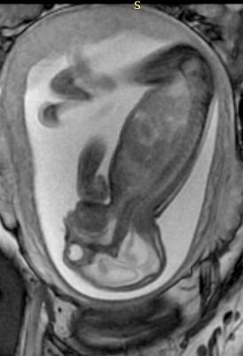
Prenatal MRI of a Zika fetus showing enlarged cerebral fluid space, dilation of the cerebral ventricles, thinning of brain tissue, poorly developed cerebellum and the absence of brain cortical gyri. (Image coutesy of RSNA)
March 15, 2021 — Magnetic resonance imaging (MRI) scanning can more precisely define and detect head, neck, thoracic, abdominal and spinal malformations in unborn babies, finds a large multidisciplinary study led by King's College London with Evelina London Children's Hospital, Great Ormond Street Hospital and UCL.
In the study, published in Lancet Child and Adolescent Health, the team of researchers and clinicians demonstrate the ways that MRI scanning can show malformations in great detail, including their effect on surrounding structures. Importantly, they note that MRI is a very safe procedure for pregnant women and their babies.
They say the work is invaluable both to clinicians caring for babies before they are born and for teams planning care of the baby after delivery.
Recent research has concentrated on correcting for fetal movement in fetal brain MRI and, more recently, for imaging the fetal heart. However, there is an increasing demand to assess the entire fetus with MRI and research from King's College London School of Biomedical Engineering & Imaging Sciences at Evelina London Children's Hospital, have recently been able to develop a post-acquisition pipeline to motion correct and volume reconstruct images of the whole fetal body.
Lead researcher, Professor Mary Rutherford, from the School of Biomedical Engineering & Imaging Sciences said ultrasound remains the gold standard for fetal screening and indeed is complementary to these optimised MRI approaches for evaluating abnormalities of the fetal body.
"Until now, ultrasound has been the modality of choice to diagnose those anomalies. However, sometimes the ability of ultrasound to define the most detailed anatomy is limited. MRI scanning offers the potential to more precisely define malformations that could help support clinicians in their planning of care and counselling of parents."
MRI is commonly used in the classification of fetal brain anomalies. Although its use in fetal body anomalies is less widely adopted, advances have led to the validation of its role in the antenatal investigation of several conditions including the investigation of fetuses with spina bifida: imaging of the fetal brain along with the spinal cord is an important factor in evaluating which patients could benefit from fetal surgery.
For fetal neck masses, MRI provides a clear advantage over conventional ultrasound for assessing tumour extension and giving a 3D visualization of the tumor's relation to the airway.
MRI may also be better than ultrasound for distinguishing between normal and abnormal lung tissue, and in making other diagnoses such as diaphragmatic hernia—particularly in late gestation, when doing so with ultrasound is challenging.
New approaches to imaging the fetal body with MRI allows both motion correction of the fetal images and volume reconstructions of body organs and defects. Researchers say this improves visualisation and therefore detection and characterisation of abnormalities.
The project brought together surgeons, fetal medicine specialists, radiologists and physicists to review the use of magnetic resonance imaging to investigate conditions in the unborn baby; this approach has already been integrated into clinical practice at Evelina London, which is part of Guy's and St Thomas' NHS Foundation Trust. Videos and images are also available for viewing.
Ongoing work is focused on a fully automated process suitable for clinical translation and wider dissemination into clinical practice.
For more information: www.

 May 17, 2024