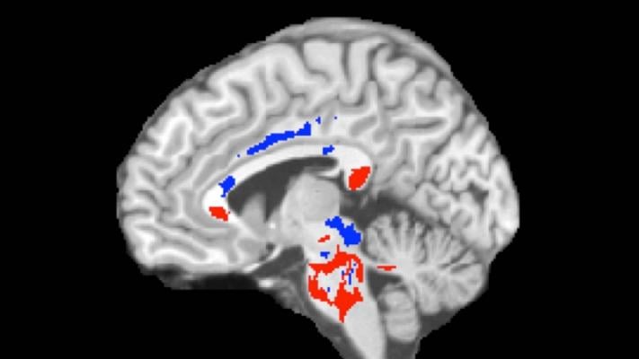
This image of a concussion patient's brain shows low FA areas (red) probably signifying injured white matter, plus high FA areas (blue) perhaps indicating more efficient white-matter connections compensating for concussion damage. A large amount of high FA predicts recovery from concussion. Image courtesy of PRNewsFoto/Albert Einstein College of Med.
June 9, 2016 — Using an advanced imaging technique, researchers at Albert Einstein College of Medicine and Montefiore Health System were able to predict which patients who'd recently suffered concussions were likely to fully recover. The study also sheds light on the brain's mechanisms for repairing or compensating for concussion injuries — information that could speed the development of therapies.
The study was published online in the American Journal of Neuroradiology.
"Our study presents for the first time a precision approach to harness imaging at the time of concussion to forecast outcomes a year later," said study leader Michael L. Lipton, M.D., Ph.D., professor of radiology, of psychiatry and behavioral sciences, and of neuroscience, as well as associate director of the Gruss Magnetic Resonance Research Center (MRRC) at Einstein and director of MRI services at Montefiore. "While we still lack effective treatments, we now have a better understanding of the neurological mechanisms that underlie a favorable response to concussion, which opens a new window on how to look at therapies and to measure their effectiveness."
Each year, 2.5 million people in the United States sustain traumatic brain injuries (TBI), according to the Centers for Disease Control and Prevention. Concussions account for at least 75 percent of these injuries. Diagnosing concussion is based on assessing symptoms; there is no objective biomarker or test. Symptoms can vary widely — lasting for just seconds or sometimes not appearing for days or weeks after injury. Common symptoms include seizures, trouble sleeping, decreased coordination, repeated vomiting or nausea, confusion and slurred speech.
"While most people think of concussions as a mild and short-lived injury, 15 to 30 percent of patients are left with symptoms that persist indefinitely," said Sara Strauss, M.D., the study's lead author and resident in the department of radiology at Montefiore. "Until now, we haven't had a reliable way to differentiate in advance those who may be burdened long-term and those who would have a complete recovery."
Conventional imaging techniques, such as computed tomography (CT) scans and magnetic resonance imaging (MRI), cannot detect the subtle damage to axons (the nerve fibers that constitute the brain's white matter) that is associated with concussions. But in a previous study, Lipton and his colleagues demonstrated that an advanced form of MRI called diffusion tensor imaging (DTI) can detect concussion-related damage to axons. It does so by "seeing" the movement of water molecules along axons, which allows researchers to measure the uniformity of water movement (called fractional anisotropy, or FA) throughout the brain. Finding a low FA brain region, for example, indicates structural damage that has impeded water movement in that area.
In the current study, Lipton tested whether brain abnormalities identified on DTI of individual concussion patients could distinguish between those patients who will eventually recover and those who will not. DTI was performed on 39 patients diagnosed with mild TBI by an emergency room physician within 16 days of the initial injury and on 40 healthy controls. The DTI image of each patient was compared with images for the entire group of healthy controls to see where patients' brains were abnormal. Patients were also assessed for three measures: cognitive function, post-concussion symptoms and health-related quality of life measures. A year later, 26 of the concussion patients returned for follow-up assessments.
DTI imaging comparing concussion patients and healthy controls revealed two types of white-matter abnormalities in patients: (1) areas of abnormally low FA (red, in associated image) that correlate with axon damage and the cognitive impairment that can affect concussion patients; and (2) other brain areas with abnormally high FA (blue) that may indicate where the brain has responded favorably to injury, perhaps by more efficiently connecting axons or by remyelinating injured tissue (i.e., forming fatty tissue around nerves, which allows nerve impulses to move more quickly).
The amount of high FA imaged in brains predicted patients' outcomes following concussion. Having a greater volume of abnormally high FA white-matter areas (perhaps indicating good compensation for concussion damage) was associated with better outcomes on follow-up assessments. (This doesn't mean that the low FA areas showing white-matter damage aren't important — just that they're not useful in predicting recovery from concussion a year later.)
"Being able to predict which patients have a good or bad prognosis has tremendous implications for discovering and evaluating treatments for concussion," said Lipton. "Developing an effective intervention requires first identifying the people who need it. Seventy to 85 percent of concussion patients get better by themselves, which makes it difficult to learn whether any treatment is actually helping. Our imaging technique allows researchers to test potential therapies on those concussion patients who can truly benefit from them."
Lipton noted that most therapies tried so far for TBI have focused on reducing damage from brain injury or preventing an injury from progressing, but none has proven effective. "Our findings," he said, "suggest that it might be worthwhile to try a different strategy — namely, attempting to enhance the brain's innate abilities to compensate functionally and structurally for whatever damage has been done."
Lipton cautions that further studies are needed to validate this approach for predicting concussion outcomes. "While we were able to predict the outcomes for the patients in our study; more refined approaches — incorporating additional patient and injury characteristics, for example — may be needed when applying the test on widely differing individuals," he said.
The study is titled, "Bidirectional Changes in Anisotropy are Associated with Outcomes in Mild Traumatic Brain Injury." The other contributors are: S. B. Strauss, Namhee Kim, Ph.D., Craig Branch, Ph.D., M.E. Kahn, Mimi Kim, Sc.D., Richard Lipton, M.D., Jennifer Provataris, M.D., H.F. Scholl and Molly Zimmerman, Ph.D., all at Einstein.
The study was funded by grants from the National Institutes of Health (NS082432-03).
The authors declare no relevant conflicts of interest.
For more information: www.ajnr.org

 April 24, 2024


