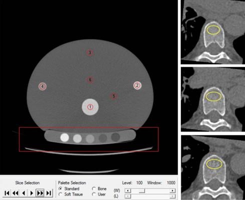
The Mindways Solid phantom with volume of interest in the quality assurance phantom (red circles, left side). A participant's noncontrast-enhanced axial CT (right side) with volume of interest (yellow circles) in the trabecular bone compartment of three vertebrae for bone mineral density measurements. Image courtesy of Radiological Society of North America
July 15, 2020 — Cardiac CT exams performed to assess heart health also provide an effective way to screen for osteoporosis, potentially speeding treatment to the previously undiagnosed, according to a study published in Radiology.
Osteoporosis is a disease that causes the bones to weaken and become vulnerable to fracture. It affects an estimated 200 million people worldwide. Early detection and treatment are important, as several classes of drugs are effective at reducing the risk of fractures that exact a devasting toll on victims. A National Osteoporosis Foundation report last year found that nearly 20 percent of Medicare fee-for-service beneficiaries died within 12 months of a new osteoporotic fracture.
Bone mineral density (BMD) tests can diagnose osteoporosis, but the number of people who get these tests is suboptimal.
"Osteoporosis is a prevalent, under-diagnosed and treatable disease associated with increased morbidity and mortality," said study lead author Josephine Therkildsen, M.D., from Herning Hospital, Hospital Unit West, in Herning, Denmark. "Effective anti-osteoporotic treatment exists and so, identifying individuals with greater fracture rate who may benefit from such treatment is imperative."
Therkildsen and colleagues recently looked at cardiac CT, a test done to assess heart health, as an opportunistic way to screen for osteoporosis. Because the cardiac CT scan also visualizes the thoracic vertebrae, the bones that form the vertebral spine in the upper trunk, it is relatively easy to add a BMD test to the procedure.
The study involved 1,487 participants who underwent cardiac CT for evaluation of heart disease. Participants also had BMD testing of three thoracic vertebrae using quantitative CT software.
Of the 1,487 people in the study, 179, or 12%, had very low BMD. During follow-up of just over three years on average, 80 of the participants, or 5.3%, were diagnosed with a fracture. The fracture was osteoporosis-related in 31 of the 80 people.
The association between a very low BMD and a higher rate of fracture strongly suggests that thoracic spine BMD may be used to guide osteoporosis preventive measures and treatment decisions, the study authors said.
Adding BMD testing to cardiac CT is feasible and applicable in a clinical setting, according to Therkildsen. It does not add time to the exam and doesn't expose the patient to any additional radiation. In fact, Therkildsen said, technological advances over time have reduced the radiation dose given at cardiac CT. BMD measurements can be made using existing non-enhanced CT images as long as a suitable calibration system is ensured, scanner stability is continuously monitored and systematic imaging acquisition techniques are implemented.
"We believe that opportunistic BMD testing using routine CT scans can be done with little change to normal clinical practice and with the benefit of identifying individuals with a greater fracture rate," Therkildsen said.
Although the researchers used cardiac CT images in the study, in theory any CT images that include a view of relevant bone structures could be used for measuring BMD. The development of fully automated software for BMD measurements further enhances the adaptability and convenience of this approach.
Additional research will help pin down the optimal BMD cut-off values for treatment while providing more data on fracture risk based on gender and areas of the body. The impact of clinical risk factors in combination with BMD measured from CT scans for the assessment of fracture risk would also benefit further study.
"Our research group is dedicated to extend the research in this field, as we believe it should be added to clinical practice," Therkildsen said.
For more information: www.rsna.org

 May 15, 2024