Angiographic imaging system vendors have developed several new technologies to address emerging cath lab trends, including the need to reduce radiation dose, improve image quality and enable advanced procedural image guidance. All three of these points have become increasingly important as more complex procedures are attempted in interventional labs and hybrid operating rooms (OR).
Advanced Imaging, Guidance, Planning
Newer imaging systems enable advanced 3-D imaging with rotational angiography, which uses a quick spin around the patient to create a computed tomography (CT)-like, 3-D image of the anatomy. This can all be done tableside in the cath lab. Some systems allow these images, or CT or magnetic resonance imaging (MRI) 3-D images, to be overlaid or fused with the live 2-D fluoroscopic images. This fusion technology is used with transcatheter aortic valve replacement (TAVR) planning and navigation software to better guide precise device placement. Software also allows 3-D images to be integrated with EP electromapping systems to guide catheter ablation procedures without the need for live fluoroscopy, helping to reduce dose.
At the Radiological Society of North America (RSNA) 2014 annual meeting, Siemens introduced its Artis with Pure technology, which simplifies the use of Siemens’ 3-D rotational angiography. The software suggests imaging protocols for ease of use and offers step-by-step instructions to create a fusion image.
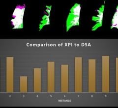
Siemens’ Syngo Dyna 4-D imaging now enables a combined digital subtraction angiography (DSA) and DynaCT (a CT-like 3-D image) at the same time to show filling of vessels with contrast. The Syngo DynaCT Smart feature can remove metal streak artifacts tableside from 3-D reconstructions. A Quick Zoom feature allows zooming in on a region of interest tableside without the need to manually manipulate images in the control room.
New software suites for various specialties have been introduced for Philips’ angiography systems, including interventional oncology, interventional neurology and for the hybrid OR suite. The oncology suite can perform two passes with the C-arm to collect images of the arterial and tumor phases with contrast to combine them into a single image to help guide embolizations. The software also features automated feeder vessel detection. Philips EchoNavigator software enables fusion imaging with live 3-D/4-D transesophageal echo (TEE).
At RSNA 2015, GE showcased its Assist brand, a collection of new interventional imaging software packages designed for specific clinical subspecialists. The suite offers simple to use fusion imaging for computed tomography (CT) 3-D image reconstructions that can be overlaid or fused with live angiography in the cath lab. The new Assist suite of targeted software solutions includes Vessel Assist to better navigate vessels for chronic total occlusions (CTO), transjugular intrahepatic portosystemic shunt (TIPS) and interventional neuroradiology arteriovenous malformation (AVM) and aneurysms. The EVAR Assist aids endovascular aortic repair (EVAR). Needle Assist is for bone interventions and pelvic bone osteosynthesis. FlightPlan for Liver helps with liver embolizations. Valve Assist is for TAVR. PCI Assist aids complex percutaneous coronary intervention (PCI) navigation.
At the 2015 American College of Cardiology (ACC) meeting, Toshiba highlighted its 3-D rotational angiography capabilities and its new transcatheter valve planning and image fusion capabilities.
Newer Systems
Siemens introduced several new systems in its Artis series, including the Artis Q, Artis Q.zen and the Artis one. These incorporate new X-ray tube and detector technology to not only lower dose, but also cut energy consumption by up to 20 percent from previous generation Artis systems. Siemens’ new X-ray tube uses flat emitter technology, which enables smaller, square focal spots that the vendor said leads to as much as 70 percent improved visibility of small vessels. The Artis Q.zen combines this X-ray tube with a new crystalline detector to enable doses as low as half the standard levels.
The Artis Q, Artis Q.zen and Artis one include the Clearstent Live application, which freezes motion in the region of a stent, allowing the physician to mask out movement of the beating heart and place the stent in precisely. The Artis one also offers new tools for cardiac imaging, including HeartSweep, which uses dual-axis rotational angiography to image the entire heart in a single, smooth C-arm movement instead of multiple acquisitions.
At RSNA 2014, Toshiba introduced its new line of combination CT/angiography systems. It combines an Infinix Elite angiography system with an Aquilion Prime or Aquilion One Vision Edition CT system. This offers an all-in-one interventional lab and CT solution to deliver real-time CT images during procedures instead of CT-like rotational angiography images. It can significantly improve workflow with its SURE Guidance technology, allowing image transition between modalities. At RSNA 2015, Toshiba showcased its Infinix 4DCT, which merges the Infinix Elite angiography system and Aquilion One Vision edition CT system. The Aquilion One Vision is capable of capturing an entire organ in one rotation with 640 slices and 16 cm of anatomical coverage.
Shimadzu’s new angiography system platform is the Trinias series. It uses high-speed Score Pro image processing technology, and a smart ergonomic design aids intuitive functionality. The Score process enhances fluoroscopic images while maintaining as low as achievable dose considerations. Trinias offers a new 12-inch Crossover flat panel detector for multi-procedure versatility. The systems provide good visibility and numerous image guidance functions, as well as 3-D applications. When used as a cardiac or neuro interventional system, Trinias is equipped with a new 8-inch flat panel detector.
GE Healthcare introduced its revolutionary Discovery IGS series in 2013. The laser-guided system captures the advantages of floor- and ceiling-mounted fixed systems and the mobility advantage of mobile C-arms without any of their inherent limitations. The imaging device is on a mobile, wheeled gantry that can be moved anywhere in the room like a mobile C-arm, but offers precision, high-quality imaging and features of a fixed system. At the touch of a button, clinicians can move the system to the table for imaging various anatomies and then move it aside and park it, allowing room for personnel and equipment during surgery. The gantry comes with a new wide-bore design, which allows for steep angles and ease in 3-D acquisition.
 May 20, 2024