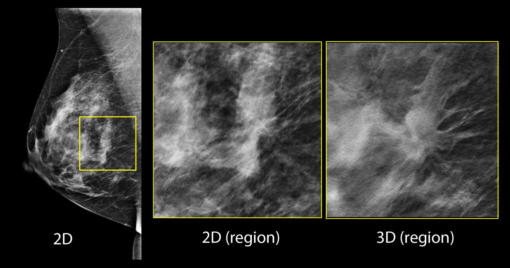
In the screening 2-D mammogram (left image), there is a possible lesion in the central breast, but its margins are difficult to assess. Using tomosynthesis this can be clearly seen to be a spiculated mass, and almost certainly a malignancy.
Just a few short years ago, the only options for breast imaging were 2-D mammography, but thanks to rapidly advancing technology to address accuracy concerns in reading dense breast images, today there several options, including 3-D tomosynthesis.
"Tomosynthesis has really become a game changer in the caring for women's breast health," said Laurie Fajardo, M.D., MBA, FACR, FSBI, chief of breast imaging, University of Iowa Hospitals and Clinics in Iowa City. "Being able to look at the breast tissue in a 3-D rendering helps us detect cancers better, and it also reduces the uncertainty of looking at a mammogram. Some of the 2-D mammograms still have a fair amount of uncertainly, particularly when women had dense breast tissue or complex breast tissue, and that meant that some of these women had to be called back for extra pictures after their screening mammogram. Not only does tomosynthesis find more cancers smaller and earlier, it also reduces the number of women who have to get called back for extra pictures that turn out not to be anything that is worrisome."
The University of Iowa Hospitals and Clinics has an outpatient imaging center that includes two tomosynthesis units, and its hospital-based center has three. "We perform tomosynthesis breast cancer screening for all women for all densities," stated Fajardo. "The literature, especially the larger Oslo Beast Cancer Screening Trial from Norway, shows us that there really is a benefit to 3-D mammography for any breast density. Tomosynthesis has been shown to perform better for breast cancer detection."
The Technology
Currently, there are three tomosynthesis systems on the market. Hologic's Selenia Dimensions mammography system offers earlier detection of breast cancers, clearer lesion images and reduction in the number of unnecessary biopsies, minimizing patient anxiety and unnecessary costs. GE's SenoClaire 3-D breast tomosynthesis offers clarity at a low dose using a short X-ray sweep around the compressed breast with only nine exposures. And most recently, Siemens received FDA approval for its Mammomat Inspiration with Tomosynthesis Option, a breast tomosynthesis add-on option for its Mammomat Inspiration digital mammography platform.
The premise of the technology is that it shoots multiple images and creates a 3-D dataset that produces multiple slices - between 40-140 - at 2 mm each, that can be rolled through and looked at in different sections. "Basically you are removing the overlying breast tissue that would be above or below the potential abnormalities, so we no longer have to deal with the possibility that small cancers or other worrisome findings in the breast will be hidden by the surrounding breast tissue that lies above or below the lesion," Fajardo explained.
By using tomosynthesis, Fajardo said her facility has been able to avoid having patients come back for extra images for something like overlapping normal breast tissue. "In our own collection of cases of cancers that you really only see on the tomosynthesis study, even knowing where it is and going back to the 2-D mammogram, you do not see that cancer," she explained. "And of those, cancers that can only be detected by tomosynthesis tend to be more aggressive; they are not low-grade cancers or indolent ductal carcinoma in situ (DCIS) cancers, so some cancers were criticized as being over-diagnosed, and slow-growing cancers and slow-growing DCIS are those kinds of cancers. Tomosynthesis has been shown not to increase the detection of those cancers."
Fajardo went on to explain that the cancers tomosynthesis finds tend to be higher developed grade cancers, typically grade 2 and grade 3. Finding those cancers earlier on a tomosynthesis study and not a 2-D study is going to help prevent them from becoming a palpable cancer before the next screening study. "The value of tomosynthesis has certainly been demonstrated in a number of single-institutional and multi-institutional trials, including the Oslo Breast Cancer Screening Trial," she stated. "I really think that tomosynthesis has been the largest breakthrough in screening technology over the last 10 years because not only does it find more cancers, it reduces the recall rate, and we've really not had a technology that does both of those things in tandem for about the last decade."
Ancillary Modalities
Emerging ancillary imaging modalities, including breast magnetic resonance imaging (MRI), have been a hot topic over the past year or so, but it is important to note that MRI still has issues with its access. "Breast MRI is a wonderful technology that has been used for many years, primarily in the diagnostic setting, and now for women who have very high risk for breast cancer - above percentages that we can measure - an insurance company will pay for them to have a screening MRI every year," Fajardo explained, adding that it is still recommended that mammography and 3-D mammography be the primary screening examination and that these other modalities be supplemental. "If MR was widely available, inexpensive, did not require contrast and performed as well as the MRI that we do with the contrast injection, it would compete very favorably with 3-D mammography. But unfortunately, access, expense, the time that it takes to do these examinations and the need to inject contrast limit its use to applying it to everybody in the U.S. population," she said.
Automated breast ultrasound screening, or ABUS, is an ultrasound examination of the entire breast, and is a supplemental screening. It is best used for women with dense breasts. "After a woman has had a mammogram, ABUS has been known to pick up additional cancers that 2-D mammography did not pick up," said Fajardo. "To date, whole-breast screening ultrasound has not been compared to 3-D or tomosynthesis. The relative disadvantages of this ultrasound examination are that you can have a very high false-positive rate. That being said, it will show things on the screening study that when you bring the patient back and do additional examination or perform a biopsy, it turns out not to be cancer. So, it finds extra cancers, but it also finds things that aren't cancer, so it has some limitations.
"We talked about tomosynthesis raising the cancer detection rate but lowering the false positive rate," she continued. "I think whole breast screening ultrasound needs to be refined to the level that it can do the same performance."
There also are nuclear approaches to breast imaging. Positron emission mammography, or PEM, uses positron emission tomography (PET) with a fluorine-18 fluorodeoxyglucose (F-18 FDG) radiotracer. Molecular breast imaging (MBI) uses single-photon emission computed tomography (SPECT) with a technetium-99 (mTc-99) sestamibi radiotracer. According to Fajardo, both of those technologies have been shown to detect breast cancers, but again they are complex, and require the injection of radioactive tracers. In addition, they are not widely available and are relatively expensive when compared to a screening mammogram or a screening tomosynthesis study.
Legislation on Breast Density
There has been a nation-wide push in legislation to make breast density reporting mandatory. "I think this movement has had some beneficial effects to women and the overall health benefits in the United States," said Fajardo. "We do know that dense breast tissue can, No. 1, hide cancers, and No. 2, because the woman with more dense breast tissue has more fibroglandular tissue in her breast, she is at a slightly increased risk of developing breast cancer compared to women who have less fibroglandular tissue and more fatty tissue in their breasts. I think this awareness has been good to educate women about this potential risk factor. And it's also been very good because it has caused physicians, mainly breast imagers, to think about what other modalities can be offered to these women. Tomosynthesis has certainly been a breakthrough for looking at dense breasts."
In addition to manually entering a BI-RADS breast density (fibroglandular tissue density) rating, some vendors offer automated density assessment technology to remove variation between readers.
Future of Imaging
The future continues to look promising for breast imaging. Five to 10 years down the road, Fajardo believes there will be a rapid uptake of 3-D mammography into the market, and that within three to five years, more than half of the mammography systems in the United States will be 3-D.
"The reason I believe that 2-D mammography will eventually be phased out is that it is possible for us today to take the 3-D dataset from a 3-D tomosynthesis mammogram and create a 2-D mammogram," Fajardo said. "So for radiologists who like to see the good old-fashioned 2-D mammogram along with their 3-D dataset, you can make the synthetic mammograms from the tomosynthesis, and you don't need to give the patient extra X-ray dose. There already is some software that is FDA approved to make synthetic mammography, and a lot of vendors are working on improving that and having their own synthetic mammography available. It is my belief that within three to five years there will be very little need to perform 2-D mammography any more."
Additional Resources on ITN for Breast Imaging and Dense Breast technology Information
VIDEO: Advances and Trends in Breast Imaging
Mammographic Breast Density — What It Means
Dense Breast Tissue: Supplemental Imaging
Breast Density: Are You Informed?

 July 09, 2025


