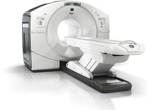
Photo courtesy of GE Healthcare
The last 12 months have seen significant growth in the positron emission tomography/computed tomography (PET/CT) segment of radiology, from both a manufacturing and a research perspective. One brand-new system received U.S. Food and Drug Administration (FDA) market clearance just months ago, while another started making its way into hospitals in the second half of last year. Several manufacturers also released software updates to help integrate PET/CT imaging into radiation therapy planning and execution. The increased interest was supported by several large studies exploring the advanced applications of PET/CT in oncology imaging.
New Systems
Siemens made the first big splash for PET/CT in 2016 when it received FDA approval for the Biograph Horizon system in January. The system is designed to offer high-end service at a lower total cost of ownership, ideal for institutions looking to replace existing PET/CT scanners. Siemens focused on keeping operations and maintenance costs down by introducing several automated features, including:
• Reconstruction scans that run alongside acquisition to deliver the image 30 seconds after the scan is completed. Scans are performed in as little as 5 minutes;
• Gentle system warm-up and automatic standby for reduced power consumption;
• Protocol-based exams such as Quanti QC, which runs quality control procedures overnight; and
• The Guardian Program, which predicts system issues before they occur. Siemens said this can lower costs related to unplanned downtime by
43 percent.
Unlike other PET/CT systems, Biograph Horizon utilizes LSO crystals, which scintillate faster and have a higher light output than BGO crystals, enabling higher image resolution and contrast as well as time-of-flight acquisition. Image processing is done through syngo.via Molecular Imaging Workplace, which offers automated prefetching, preprocessing, and display and comparison of previous findings.
In addition to Biograph Horizon, Siemens launched several PET/CT enhancements for radiation therapy at the 2015 American Society for Radiation Oncology (ASTRO) annual meeting last October. Perhaps most significant was the RT Pro edition of the Biograph mCT family, specifically designed for radiation therapy treatment planning. The new edition employs an advanced algorithm and metal artifact reduction for better tumor delineation, and motion management technologies like FlowMotion continuous bed motion technology for CT-like organ-focused acquisition. Thanks to a partnership with Varian Medical Systems, all Siemens PET/CT systems now include respiratory gating for scanners (RGSC) technology to help synchronize the workflow between diagnostic imaging and respiratory-gated treatment delivery.
The Biograph 64 mCT Flow also got a software update in 2015 when Siemens released the latest version of VG60, which improves image quality to help streamline oncology imaging services with radiation therapy. New features include removal of metal artifacts, improved large field-of-view imaging and respiratory motion correction.
Meanwhile, Toshiba saw the first U.S. installs of its debut PET/CT system, Celesteion, in the back half of 2015 following FDA approval the previous year. The company said it offers the industry’s largest bore (90 cm CT and 88 cm PET) and widest field of view, plus Toshiba’s AIDR 3-D (Adaptive Iterative Dose Reconstruction) dose-lowering technology. The system is ideal for tumor detection, treatment evaluation and CT simulation.
Philips introduced digital PET detectors to the PET/CT market when it unveiled the Vereos at the Radiological Society of North America (RSNA) 2013 annual meeting. The system uses Philips’ digital photon counting technology in its digital silicon detectors, allowing improved image clarity over traditional analog photomultipliers. Philips said this results in a step change in performance that approximately doubles sensitivity gain, volumetric resolution and quantitative accuracy compared to analog systems. These radical improvements can ultimately be translated into high image quality, increased diagnostic confidence, improved treatment planning and faster workflows.
GE Healthcare highlighted its Discovery IQ PET/CT system, which became FDA-cleared in September 2014, at RSNA 2015. It enables both high image quality and intelligent quantitation. GE said it delivers the highest NEMA (National Electrical Manufacturers Association) sensitivity (up to 22 cps/kBq) and the largest axial field-of-view (up to 26 cm) compared to other PET/CT equipment.
PET/CT in Cancer Detection Studies
While PET/CT already has numerous clinical applications for oncology imaging, several recent studies have uncovered new ways to take advantage of the technology.
A pair of studies found PET/CT is particularly beneficial for detection of prostate cancer. The earlier study, published in the July 2015 issue of The Journal of Nuclear Medicine, found that PET/CT, with the aid of a PSMA biomarker, was more specific than magnetic resonance imaging (MRI) in detecting clinically significant high-grade prostate cancer lesions; the biomarker was also able to better distinguish benign prostate lesions from primary prostate cancer.
The second study, conducted by researchers from Thomas Jefferson University and published in the January 2016 issue of Urology, concluded that PET/CT more accurately detects prostate cancer than any other test available — radiographical, biopsy or blood. They examined 25 patients who had opted for radical prostatectomy using PET/CT with a specially developed intravenous biomarker that attaches to cancerous prostate cells. The agent was able to identify not only 105 of the 107 lesions found in pathological exams of the removed glands, but an additional nine lesions not found pathologically.
One study out of the United Kingdom, published in the New England Journal of Medicine, found that PET/CT was better at detecting head and neck cancer cells remaining after primary chemoradiotherapy — which happens for 20 percent of patients — than traditional neck dissection surgery. Researchers looked at 564 cases across 37 centers in a five-year period; half were randomized to image-guided surveillance and half to the neck dissection control group. Thanks to PET/CT, only 19 percent of the image-guided surveillance patients needed to have invasive neck dissection surgery, compared to 78 percent in the control group.

 December 10, 2025