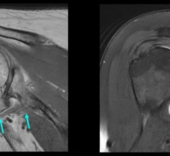
Image courtesy of Spectrum Health
June 30, 2015 - Congenital heart experts from Spectrum Health Helen DeVos Children's Hospital have successfully integrated two common imaging techniques to produce a three-dimensional anatomic model of a patient's heart.
The 3-D model printing of patients' hearts has become more common in recent years as part of an emerging, experimental field devoted to enhanced visualization of individual cardiac structures and characteristics. But this is the first time the integration of computed tomography (CT) and three-dimensional transesophageal echocardiography (TEE) has successfully been used for printing a hybrid 3-D model of a patient's heart. A proof-of-concept study authored by the Spectrum Health experts also opens the way for these techniques to be used in combination with a third tool – magnetic resonance imaging (MRI).
"Hybrid 3-D printing integrates the best aspects of two or more imaging modalities, which can potentially enhance diagnosis, as well as interventional and surgical planning," said Jordan Gosnell, Helen DeVos Children's Hospital cardiac sonographer, and lead author of the study. "Previous methods of 3-D printing utilize only one imaging modality, which may not be as accurate as merging two or more datasets."
The team used specialized software to register images from the two imaging modalities to selectively integrate datasets to produce an accurate anatomic model of the heart. The result creates more detailed and anatomically accurate 3-D renderings and printed models, which may enable physicians to better diagnose and treat heart disease.
CT and MRI are established imaging tools for producing 3-D printable models. 3-D TEE recently was reported by Joseph Vettukattil, M.D., and his Helen DeVos Children's Hospital colleagues to be a feasible imaging technique to generate 3-D printing in congenital heart disease. Vettukattil is co-director of the Helen DeVos Children's Hospital Congenital Heart Center, division chief, pediatric cardiology, and senior author of the study.
According to Vettukattil and his colleagues, each imaging tool has different strengths, which can improve and enhance 3-D printing:
- CT enhances visualization of the outside anatomy of the heart;
- MRI is superior to other imaging techniques for measuring the interior of the heart, including the right and left ventricles or main chambers of the heart, as well as the heart's muscular tissue; and
- 3-D TEE provides the best visualization of valve anatomy.
"This is a huge leap for individualized medicine in cardiology and congenital heart disease," said Vettukattil. "The technology could be beneficial to cardiologists and surgeons. The model will promote better diagnostic capability and improved interventional and surgical planning, which will help determine whether a condition can be treated via transcatheter route or if it requires surgery."
Vettukattil is known internationally for his work and research with three- and four-dimensional echocardiography. Most notably, Vettukattil developed the advanced technique of multiplanar reformatting in echocardiography, a method used to slice heart structures in infinite planes through the three dimensions in a virtual environment similar to a cardiac pathologist dissecting the heart to reveal underlying pathology. Commonly used with other diagnostic technologies, such as CTs, Vettukattil pioneered its use in echocardiography to evaluate complex heart defects.
Vettukattil is presenting the findings of the proof-of-concept study June 24-27 at the CSI 2015 - Catheter Interventions in Congenital, Structural and Valvular Heart Diseases Congress in Frankfurt, Germany. The presentation will demonstrate the feasibility of printing 3-D cardiovascular models derived from multiple imaging modalities.
The Helen DeVos Children's Hospital team worked with the Mimics Innovation Suite software from Materialise, which printed the model using its HeartPrint Flex technology. Gosnell worked on integration of the imaging modalities, collaborating with Materialise's U.S. headquarters in Plymouth, Michigan, to produce the final 3-D rendering. Vettukattil devised the concept of integrating two or more imaging modalities for 3-D printing.
Further research is required to evaluate the efficacy of hybrid 3-D models in decision-making for transcatheter or surgical interventions.
For more information: www.spectrumhealth.org
 May 17, 2024