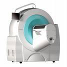
Siemens Inveon PET
April 22, 2010 - Researchers came one step closer to improving the selection of patients for targeted breast cancer therapies.
At the 101st meeting of the American Association of Cancer Research in Washington, D.C., a research team at Radboud University Nijmegen Medical Center, Nijmegen, The Netherlands on April 19 presented results from a study in which they used in vivo PET (positron emission tomography) and SPECT (single photo emission computed tomography) molecular imaging techniques in animal subjects with triple negative breast cancer. The data shows this facilitated patient selection for targeted therapies.
The purpose of the study was to develop a noninvasive method of visualizing the insulin-like growth factor 1 receptor, IGF-1R, which is expressed by 30 percent to 40 percent of patients with triple negative breast cancer. This makes them uniquely identifiable and able to be treated with IGF-1R antibodies.
“The use of molecular imaging techniques, such as PET and SPECT, has been helpful in the progression of our research toward finding molecular targets to visualize the IGF-1R, expressed by cells of patients with triple negative breast cancer. In the future, the ability to visualize the receptor may enable more effective patient selection from the triple negative breast cancer patient population for IGF-1R targeted therapy,” said Otto C. Boerman, of the department of Nuclear Medicine, Radboud University Medical Center, Nijmegen.
Siemens Inveon PET and MILabs U-SPECT preclinical imagers were used in the in vivo portion of the study. Small animal PET, SPECT and CT molecular imaging provides quantitative insight into understanding tumor biology and can aid in the development of therapeutic options.
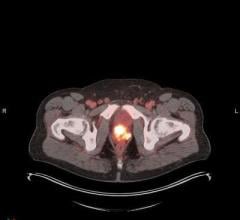
The researchers concluded that 111In-R1507 SPECT and 89Zr-R1507 PET are excellent methods to visualize IGF-1R expression in vivo in triple negative breast cancer xenografts.
The study results verified that 111In-R1507 was slowly internalized by SUM149 cells and was specifically and efficiently accumulated in the SUM149 xenografts. The specific tumor uptake was reported at 20 percent ID/g, 33 percent ID/g and 31 percent ID/g at one, three and seven days post injection, respectively. At the optimal dose for imaging, the tumor uptake was 35 percent ID/g and enabled clear visualization of the subcutaneous xenograft with increasing contrast at later time points.
The study was conducted using R1507, a monoclonal antibody directed against an epitope on the extracellular domain of the IGF-1R. It was radiolabeled with 111 Indium (111In), 125 Iodine (125I) and 89 Zirconium (89Zr). In vitro, the affinity and internalization kinetics of 111In-R1507 were determined using the IGF-1R expressing breast cancer cell line SUM149, which is an estrogen receptor, progesterone receptor and HER2 receptor negative.
In vivo studies were performed in BALB/c nude mice with subcutaneous SUM149 xenografts. To determine the pharmacodynamics of R1507, mice received an intravenous injection of 111In-R1507 and 125I-R1507. One, three and seven days post injection, the ex vivo tumor uptake was measured. Non-specific uptake was determined by coinjection of an excess unlabeled R1507. A dose escalation study was performed with 111In-R1507 to determine the optimal protein dose of R1507 for in vivo imaging (dose range 1 - 1,000 µg).
Finally, PET and SPECT images were acquired of mice with subcutaneous SUM149 tumors one, three and seven days post injection of 89Zr-R1507 and 111In-R1507, respectively.
For more information: www.siemens.com/healthcare
 May 08, 2024