August 23, 2016 — A novel magnetic resonance imaging (MRI) method that detects low levels of zinc ion can help distinguish healthy prostate tissue from cancer, UT Southwestern Medical Center radiologists have determined.
Typical MRIs do not reliably distinguish between zinc levels in healthy, malignant and benign hyperplastic prostate tissue, so discovery of the technique could eventually prove useful as a biomarker to track the progression of prostate cancer, according to researchers with the Advanced Imaging Research Center, part of UT Southwestern’s Harold C. Simmons Comprehensive Cancer Center.
“This research provides the basis for differentiating healthy prostate from prostate cancer by use of a novel Zn(II) ion sensing molecule and MRI,” said senior author A. Dean Sherry, M.D., director of the Advanced Imaging Research Center and professor of radiology at UT Southwestern.
The findings appear in the Proceedings of the National Academy of Sciences.
“The potential for translating this method to human clinical imaging is very good, and will be useful for diagnostic purposes. The method may prove useful for monitoring therapies used to treat prostate cancer,” said Sherry, who is also professor of chemistry at UT Dallas, where he holds the Cecil and Ida Green Distinguished Chair in Systems Biology.
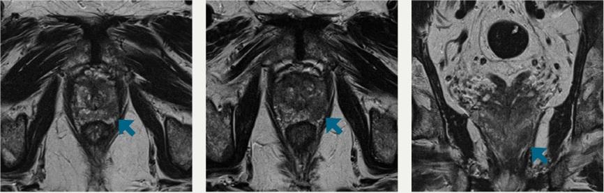
The majority of prostate cancers are classified as adenocarcinomas and originate in epithelial cells. The UTSW researchers initially determined that glucose stimulates release of the zinc ions from inside epithelial cells, which they could then track on MRIs. The prostate cancer tissue secreted lower levels of zinc ions, offering an opportunity to distinguish between malignant and healthy tissue. When they tested the technique on mouse models, they were able to successfully detect small malignant lesions as early as 11 weeks, making the non-invasive imaging procedure a potentially useful method for detecting the disease and its progression.
“Prostate cancer often has no early symptoms, so identifying potential new diagnostic methods that might catch the cancer at an earlier stage or allow us to track how it is progressing is an important opportunity,” said co-author Neil Rofsky, M.D., chairman of radiology, director of translational research for the Advanced Imaging Research Center, and holder of the Effie and Wofford Cain Distinguished Chair in Diagnostic Imaging.
Prostate cancer is the most common cancer in men in the United States, after skin cancer, and is the second leading cause of death from cancer in men, according to the National Cancer Institute. Prostate cancer occurs more often in African-American men, who are more likely to die from the disease.
MRI, which uses only harmless magnetic fields and radio waves, is one of the most benign technologies in medicine for studying and diagnosing medical disorders, enabling researchers to view diseases that afflict millions of people, without the need for surgery, X-rays or radioactive tracers.
Support for this latest research came from grants from the National Institutes of Health, National Cancer Institute through the Harold C. Simmons Comprehensive Cancer Center, the American Diabetes Association, and the Robert A. Welch Foundation.
Other UTSW researchers from the AIRC, Radiology, Cell Biology, Biochemistry, Pathology, Urology and the UT Southwestern Graduate School of Biomedical Sciences included lead author Dr. Veronica Clavijo Jordan; postdoctoral researchers Dr. Su-Tang Lo, Dr. Cristian Preihs, Dr. Shanrong Zhang, and fellow Dr. Shiuhwei Chen; Dr. Payal Kapur, associate professor of pathology and urology; and Dr. Wen-Hong Li, associate professor of cell biology and biochemistry, and Southwestern Medical Foundation Scholar in Medical Research.
For more information: www.pnas.org

 April 24, 2024


