A new wave of smaller, less expensive, and portable MRI systems promises to expand the delivery of health care and the capabilities of medicine
July 18, 2023 — Magnetic resonance imaging (MRI) machines can clearly view non-bony parts of the body — soft tissue such as the brain, muscles and ligaments — as well as detect tumors, making it possible to diagnose many diseases and other conditions. However, the powerful magnets in conventional MRI machines make them expensive and bulky, confining them mainly to hospitals and other large facilities.
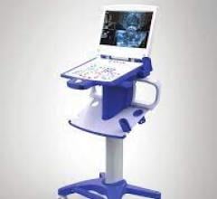
As an alternative solution, companies are developing new portable versions that have lower-strength magnetic fields. These new models can potentially expand the ways in which MRI is used. For instance, low-field MRI systems could be deployed in ambulances and other mobile settings. They also could cost much less, promising to make MRI more widely available, including in underserved communities and developing nations.
But for low-field MRI scanners to reach their full potential, more research is needed to understand the relationship between low-field images and the underlying tissue properties they represent. Researchers at the National Institute of Standards and Technology (NIST) have been working on several fronts to advance low-field MRI technology and validate methods for creating images with weaker magnetic fields.
“Magnetic resonance images of tissue differ depending on magnetic strength,” said NIST electrical engineer Kalina Jordanova. “With low-field MRI systems, the contrast of the images is different, so we need to know how human tissue looks at these lower field strengths.”
Toward these ends, researchers measured the properties of brain tissue at low magnetic field strength. Their results were published in the journal Magnetic Resonance Materials in Physics, Biology and Medicine.
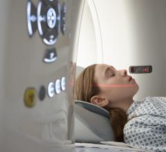
The researchers used a commercially available portable MRI machine to image brain tissue in five male and five female volunteers. The images were created using a magnetic field strength of 64 millitesla, which is at least 20 times lower than the magnetic field in conventional MRI scanners.
They collected images of the entire brain and obtained data on its gray matter (which has a high concentration of nerve cells), white matter (deeper tissues of the brain that house nerve fibers), and cerebrospinal fluid (clear fluid surrounding the brain and spinal cord).
These three brain constituents respond to the low magnetic field in different ways and produce distinctive signals that reflect their unique properties, enabling the MRI system to produce images that contain quantitative information about each constituent. “Knowing the quantitative properties of tissue allows us to develop new image collection strategies for this MRI system,” said NIST biomedical engineer Katy Keenan.
In separate work, NIST researchers are exploring several candidate materials that can significantly boost image quality in low-field MRI scans.
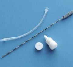
MRI contrast agents — magnetic materials that are injected into patients and enhance image contrast — make it easier for radiologists to identify anatomical features or evidence of disease and are routinely used in MRI at conventional magnetic field strengths. However, researchers are just starting to understand how contrast agents might be used with the new low-field MRI scanners. At the lower field strengths of these scanners, contrast agents may act differently than at higher field strengths, creating opportunities to use new types of magnetic materials for image enhancement.
NIST scientists and their colleagues compared the sensitivity of several magnetic contrast agents in low magnetic fields. The researchers found that iron oxide nanoparticles outperformed traditional contrast agents, which are made of the element gadolinium — a rare-earth metal. At low magnetic field strength, the nanoparticles provided good contrast using a concentration of only about one-ninth that of the gadolinium particles.
Iron oxide nanoparticles also offer the advantage that they are broken down by the human body instead of potentially accumulating in tissue, noted NIST researcher Samuel Oberdick. By comparison, a small amount of gadolinium may accumulate in tissue and could confound the interpretation of future MRI scans if it is not taken into account.
NIST researchers collaborated with the University of Florence in Italy and Hyperfine Inc. in Guilford, Connecticut, and reported their findings in the journal Scientific Reports.
For more information: https://www.nist.gov/

 May 03, 2024