Sandro Santagata, Ludwig Harvard. Image courtesy of Ludwig Cancer Research
July 3, 2023 — Researchers at the Ludwig Center at Harvard have developed a platform technology for imaging that enables integration of the methods of microscopic analysis long employed in pathology laboratories with the visualization of multiple molecular markers in individual cells that is now rapidly advancing in research labs. The latter capability, known as “multiplex” imaging, promises to revolutionize cancer diagnostics by exposing molecular traits associated with such things as a cancer’s aggressiveness or its potential vulnerability to therapy. It has, however, remained largely confined to academic labs because its methods tend to be incompatible with the standardized processes of tissue processing and staining employed for diagnosis in clinical pathology.
In the current issue of Nature Cancer, researchers led by Ludwig Harvard’s Sandro Santagata, Peter Sorger, Jia-Ren Lin and Yu-An Chen report the development of a platform, named Orion, that bridges these parallel worlds of microscopic analysis. They also validate the platform, demonstrating its use by both human experts and artificial intelligence algorithms in identifying cellular and molecular features in tumors that predict the progression-free survival of colon cancer patients.
“What this all boils down to is that Orion is a tool designed for practical use in real-life clinical settings,” said Santagata. “While the platform can certainly benefit basic cancer research, its primary focus is to enhance the diagnosis of human diseases. That has been our goal since the beginning.”
Clinical pathology relies on the microscopic analysis of tissues embedded in wax, sliced into strips about a fifteenth the width of a human hair and stained with hematoxylin and eosin (H&E) to expose the fine morphology of cells. Today, researchers are increasingly applying machine learning and artificial intelligence algorithms to the pink-and-purple images of H&E-stained tissues. Such work—which is further enriched by the ability to draw from millions of archived H&E images and samples—identifies patterns not readily apparent to the human eye, revealing information that can help improve the diagnosis and management of cancer therapy.
Meanwhile, the use of antibodies to capture molecular characteristics of tumors has evolved rapidly over the past four decades and is now indispensable to cancer diagnostics. More recently, researchers have begun simultaneously applying scores of antibodies to tissues, linking “immunofluorescence” images generated this way to gene expression in cells to generate richly textured portraits of tumors.
“With the molecular information we gather, we can identify distinct cell types, determine their current state, understand their neighboring cells and assess how they may be interacting to promote tumor growth,” Santagata explained.
A couple of issues have, however, limited the application of multiplex imaging in the clinic. First, its methods do not generally permit the evaluation of whole slides—a requirement of clinical diagnostics. Nor are they compatible with those of H&E microscopy, the workhorse of clinical pathology.
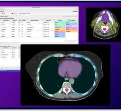
“Our platform paves the way for bridging the domains of H&E diagnostics and multiplexed tissue imaging,” said Santagata. “We have developed techniques to combine multiple antibody images with H&E staining of the same tissue section, enabling the integration of information from each source into a comprehensive view across the entire slide.”
The Ludwig Harvard team partnered with a Seattle, WA, startup named RareCyte and drew additionally on support from the Ludwig Tumor Atlas project and a Small Business Innovation Research grant from the National Cancer Institute to develop a new microscope and processes at the heart of the Orion platform.
To test the platform, the researchers analyzed colorectal cancer specimens from 40 patients to find molecular features most closely associated with bad outcomes. Sifting through some 15,000 combinations of biomarkers, they identified those most tightly linked to patient prognosis and applied them to samples from 34 other colorectal cancer patients whose outcomes were known. The researchers show that their biomarkers predicted with high accuracy—just a 1 in 20 chance of being wrong—the likelihood of poor prognosis.
Notably, combining multiplex and H&E imaging revealed relationships between the molecular markers, cell morphology and tumor topography that were illuminating. One such finding indicated that inflammation—or immune activity—at the rim of the tumor is of pathological significance. Another revealed the molecular basis of a tissue morphology associated with propensity for metastasis.
“Cancer cells typically grow together in sheets, but sometimes they lose their ability to stick together, resulting in the release of fragmented clusters. Malfunctions in the machinery of cellular adhesion can make cancer cells more invasive,” explained Santagata. “During our analysis, we saw that a critical marker of cell adhesion was decreased in tumors displaying invasive characteristics, and this reduction was associated with unfavorable outcomes.”
The researchers now plan to continue establishing proof of concept for their platform, exploring markers of such things as treatment response and resistance. Ultimately, the discoveries made using Orion will have to be validated in large clinical trials.
“The prospect of incorporating high-powered advanced imaging tools to analyze routine clinical samples is very exciting,” said Santagata. “It promises to unlock a wealth of knowledge about cancer tissues.”
For more information: [email protected]

 May 03, 2024