July 28, 2008 – Scientists have captured images of brain lesions similar to those found in Alzheimer’s disease using clinical-grade MRI in an animal model of the disease for the first time, according to research reported today at the Alzheimer's Association's 2008 International Conference on Alzheimer's Disease (ICAD 2008), in Chicago.
These lesions, known as amyloid plaques, have been “imaged” previously using very high power MRI scanners that are only used on animals, and also with PET scans combined with specialized marker chemicals. This is the first time images of plaques were captured with conventional, clinical strength MRI, according to the Alzheimer’s Association.
Two other studies reported at ICAD 2008 use MRI and advanced computer analysis also identify Alzheimer's earlier, perhaps even before symptoms are evident.
“As we get closer to the development of therapies that can slow or even stop the progression of Alzheimer’s, earlier detection of the disease becomes crucial for early intervention," said William Thies, Ph.D., vice president of medical and scientific relations at the Alzheimer’s Association. “Early evaluation and diagnosis is also important because some Alzheimer’s-like symptoms can be reversed if they are caused by treatable conditions, such as depression, drug interaction or thyroid problems.”
“As we search for ways to identify Alzheimer's early, these MRI studies show that researchers are moving closer to accurate early detection of the disease, and that we may soon be able to use this technology to determine who is at greater risk,” he added.
Definitive diagnosis of Alzheimer’s currently happens at autopsy by demonstrating the presence of characteristic brain lesions, including amyloid plaques. The ability to noninvasively show amyloid plaque levels in living people could markedly improve the diagnosis and treatment of people with Alzheimer’s.
John Ronald, a Ph.D. candidate in Medical Biophysics, along with Brian Rutt, Ph.D., and colleagues at the Robarts Research Institute and University of Western Ontario, London, Ontario, Canada, used clinical strength MRI scanners to take brain images from rabbits that had been fed a high cholesterol diet for more than two years. These animals form amyloid plaques in their brains.
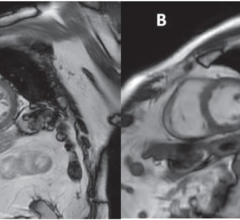
According to the researchers, the MRI scans revealed distinct signal voids - black spots - in several brain areas including the hippocampus, which is very important for memory. Autopsy examination revealed that the void areas reflected the presence of small clusters of amyloid plaques. Each cluster had high levels of iron, which the researchers say caused the MRI signal voids. These signal voids were not found in animals fed a normal diet.
“Although some of the technology used to generate these images was designed specifically for rabbits, this preliminary discovery hints at the promise of using clinical MRI scanners to visualize plaques in people with Alzheimer’s," Ronald said. “Extension of these technologies to living animals is practical, and should allow us to study the course of Alzheimer’s in animals over time.
"We have customized this MRI scanner in very important ways for microimaging. Particularly, we have added special hardware that allows the scanner to clearly detect structures smaller than 50 microns (0.05 mm) and to more sensitively detect iron-containing structures than has been possible using clinical MRI in the past,” Ronald added.
In addition to amyloid plaques, there is another brain lesion that is characteristic of Alzheimer’s - neurofibrillary tangles. The “gold standard” for post mortem measurement of Alzheimer's tangle severity is known as Braak staging.
Prashanthi Vemuri, Ph.D., Clifford R. Jack, M.D. and colleagues at the Mayo Clinic, Rochester, MN, sought to validate the ability of a new MRI analysis algorithm - known as the STructural Abnormality iNDex (STAND) score - to capture Alzheimer’s-related tangle severity. This was done by comparing the person's STAND-score derived from an MRI scan taken before death with the person’s Braak stage calculated after death.
The researchers developed an algorithm that extracts atrophy information from an individual patient’s three-dimensional MRI scan. A STAND-score is assigned based on comparing the degree of atrophy in the person's brain to atrophy patterns extracted from a large library of 160 Alzheimer’s and 160 cognitively normal subjects’ MRI scans. The STAND score is positive if the brain looks more Alzheimer's-like and negative if the scan looks normal, and can be adjusted for demographics. According to the researchers, STAND-scores have 90 percent accuracy in distinguishing the MRI scans of people with Alzheimer's from normal MRI scans.
In order to verify the adjusted STAND scores against Braak staging, the researchers identified 101 patients who had an MRI scan within four years of the time of death as well as postmortem Braak staging. They compared the two and, on a scale of 0 to 1, found the strength of association between the STAND-score and Braak stage to be 0.63 (p
In previously reported research, Christos Davatzikos, Ph.D., of the department of radiology at the University of Pennsylvania, Philadelphia; Susan Resnick, Ph.D., of the National Institute on Aging, Bethesda, M.D. and colleagues used a new computer-based image analysis technique (advanced high-dimensional pattern classification) to analyze MRI scans from participants in the Alzheimer’s Disease Neuroimaging Initiative (ADNI) and to derive an index of Alzheimer-like brain abnormality.
In a new study reported at ICAD 2008, they examined the presence and progression of such patterns in elderly individuals who are cognitively normal, and a small group with mild cognitive impairment (MCI), from the Baltimore Longitudinal Study of Aging (BLSA).
One hundred nine (109) healthy elderly participants from the BLSA were analyzed over periods of up to nine years. The researchers found that Alzheimer’s-like patterns of brain atrophy were increasingly present in participants over age 80. The rate of progression of the index of Alzheimer- like patterns over time also was higher in the older participants. Memory performance of healthy elderly and those with MCI who displayed Alzheimer-like brain atrophy patterns, as measured using standard neuropsychological batteries, was lower compared to people that didn't display these patterns and had lower Alzheimer-like index values.
“Although the clinical significance of these Alzheimer's-like patterns of brain atrophy must be further evaluated, we are very hopeful that these pattern analysis tools will provide early indicators of brain changes that resemble those seen in people with Alzheimer’s, years before memory problems are recognized clinically," Dr. Davatzikos said.
For more information: www.alz.org

 April 24, 2024