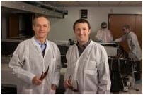
Physics Professor Donald Umstadter and doctoral student Nathan Powers in the Diocles Extreme Light Laboratory.
Using a compact but powerful laser, a research team at the University of Nebraska-Lincoln has developed a new way to generate synchrotron X-rays.
Although the high quality of synchrotron X-rays make them ideal for research ranging from the structure of matter to advanced medical images, access to the technology has been limited until now. Most traditional synchrotron X-ray devices are gigantic and costly, available only at a few sites around the world.
As reported in this week's issue of the top-ranked optics journal Nature Photonics, researchers at UNL's Extreme Light Laboratory developed a novel method to generate research-quality X-rays using a "tabletop" laser.
"Our hope is that this new technology will lead to applications that benefit both science and society," said Nathan Powers, a Ph.D. student and first author of the journal article.
Physics professor Donald Umstadter, director of the Extreme Light Laboratory, led the research project. He compared the synchrotron X-ray breakthrough to the development of personal computers, giving more people access to computing power once available only via large and costly mainframe computers. Shrinking components of advanced laser-based technology will increase the feasibility of producing high-quality X-rays in medical and university research laboratories, which in turn could lead to new applications for the X-rays.
Because the new X-ray device could be small enough to fit in a hospital or on a truck, it could lead to more widespread applications for advanced X-ray technology, UNL scientists said. New applications might include Homeland Security detecting nuclear materials concealed within a shielded container; doctors finding cancerous tumors at earlier stages; or scientists studying extremely fast reactions that occur too rapidly for observation with conventional X-rays.
Ever since synchrotron X-ray light sources were developed more than 60 years ago, they have grown in size. Some now equal the size of a college campus, with a cost in the hundreds of millions of dollars. These huge machines continue to be built, most recently in Australia and Brazil.
Like supercomputers, they provide scientists with the most advanced research capabilities, yet they are not feasible for most practical applications. Though synchrotron X-rays result in lower doses of radiation as well as high-quality images, the tens of thousands of compact X-ray devices currently in operation in hospitals or at ports worldwide produce lower quality X-rays.
In traditional synchrotron machines, electrons are accelerated to extremely high energy and then made to change direction periodically, leading them to emit energy at X-ray wavelength. At the European Synchrotron Radiation Facility in Grenoble, France, the electrons circle near the speed of light in a storage ring of 844 meters in circumference. Magnets are used to change the direction of the electrons and produce X-rays.
Pursuing an alternative approach in the recent experiments, the UNL team replaced both the electron accelerator and the magnets with laser light. They first focused their laser beam onto a gas jet, creating a beam of relativistic electrons. They then focused another laser beam onto the accelerated electron beam. This rapidly vibrated the electrons, which in turn caused them to emit a bright burst of synchrotron X-rays—a process referred to as Compton scattering. Remarkably, the light's photon energy was increased during this process by a million-fold. And yet, the combined length of the accelerator and synchrotron was less than the size of a dime.
"The X-rays that were previously generated with compact lasers lacked several of the distinguishing characteristics of synchrotron light, such as a relatively pure and tunable color spectrum, " Umstadter said. "Instead, those X-rays resembled the 'white light' emitted by the sun."
The new laser-driven device produces X-rays over a much larger range of photon energies, extending to the energy of nuclear gamma rays. Even fewer conventional synchrotron X-ray sources are capable of producing such high photon energy. Key to the breakthrough was finding a way to collide the two micro-thin beams—the scattering laser beam and the laser-accelerated electron beam.
"Our aim and timing needed to be as good as that of two sharpshooters attempting to collide their bullets in midair," Umstadter said. "Colliding our 'bullets' might have even been harder, since they travel at nearly the speed of light."

 April 09, 2024