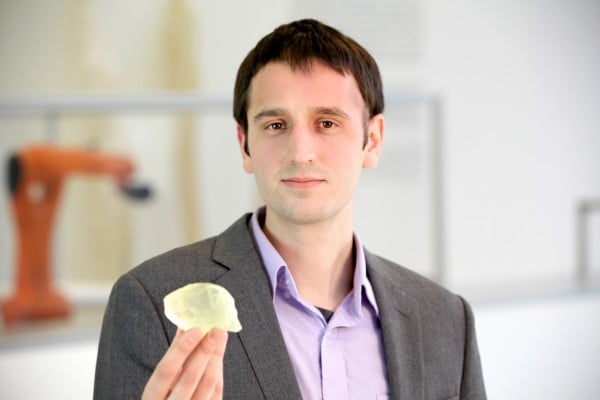
MIT graduate student Steven Keating holds a 3-D-printed copy of his cancerous astrocytoma brain tumor. It was printed by Keating with data from Brigham and Women's Hospital. Image courtesy of Paula Aguilera and Jonathan William.
An MIT graduate student’s research into 3-D printing helped doctors remove a tumor from his brain last August, and he is now sharing his story as an advocate for increased patient access to their own medical records.
In 2007, Steven Keating had his brain scanned out of sheer curiosity. Keating had joined a research study that included a magnetic resonance imaging (MRI) scan, and he asked that the scan’s raw data be returned to him. The scan revealed only a slight abnormality, near his brain’s smell center, which he was advised to have re-evaluated in a few years. A second scan, in 2010, showed no change, suggesting that the abnormality was most likely benign.
While the second scan provided reassurance, Keating’s knowledge of the abnormality — as a result of having access to the raw data from these scans — ultimately led to the detection of a baseball-sized tumor that was removed in August 2014.
Now a graduate student in the Department of Mechanical Engineering and based at the MIT Media Lab, Keating says that his curiosity saved his life — and that his experience with cancer has fueled a strong interest in advocating for open health data.
Last July, Keating noticed that he was experiencing a phantom vinegar smell for about 30 seconds every day. Knowing that his 2007 and 2010 research scans showed an abnormality near his smell center, he requested an MRI scan through MIT Medical. The scan revealed that the abnormality had grown into a tumor that needed to be removed as soon as possible.
Keating went to Brigham and Women’s Hospital (BWH) in Boston on Aug. 19 for surgery, performed by neurosurgeon E. Antonio Chiocca. Keating, though sedated, was kept awake while the tumor was removed. This was so doctors could ask him questions while they were probing and cutting brain tissue to ensure they were not damaging the brain’s language center. The 10-hour surgery was captured on video, which, at Keating’s request, was shared with him.
His recovery was quick: Keating was out of the hospital after two days, and he was back on the MIT campus within a week.
A tissue biopsy confirmed that his tumor was an IDH1-mutant malignant astrocytoma. In this type of brain cancer, the mutated IDH enzyme leads to the production of 2HG, a novel, oncogenic metabolite.
Through the Bridge Project — a collaboration between MIT’s Koch Institute for Integrative Cancer Research and the Dana-Farber/Harvard Cancer Center — a cross-institutional research team is exploring how to use 2HG as a biomarker to detect and monitor IDH-mutant cancers.
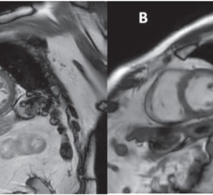
Ovidiu Andronesi, a radiologist at Massachusetts General Hospital (MGH) and a collaborator on this research, applied this monitoring technology via MRI spectroscopy imaging to scan Keating’s brain before and after his surgery. These scans show the reduction of 2HG after doctors removed the tumor; the scans were also shared with Keating, at his request.
“As a cancer scientist, hearing Steven talk about 2HG spectroscopy screening as part of his clinical care is remarkable,” says Matthew Vander Heiden, the Eisen and Chang Career Development Associate Professor of Biology and a member of the Koch Institute, who is a leader on this research project. “IDH’s role in these cancers was only discovered six years ago, and it is incredible, as well as humbling, that Steven could benefit from some of the basic science done in this short time period since IDH mutations were recognized.”
With this abundance of data, Keating is able to apply his own research interests to develop an intimate understanding of his brain and his tumor. In Oxman’s Mediated Matter Group, Keating’s research explores how to leverage 3-D printing and other fabrication methods to print everything from living organisms to entire buildings. With the resources available to him at the Media Lab, he and colleagues James Weaver and Ahmed Hosny at Harvard University’s Wyss Institute for Biologically Inspired Engineering have pored over his health data and created digital and 3-D-printed models of his tumor, brain and surgically repaired skull.
Given his up-close-and-personal experience with his health, Keating says he is now a strong believer in open sourcing and allowing patients to have easy access to their own health data. He says he was fortunate that his doctors were willing to share his data, but he did notice many small barriers along the way.
“My doctors are incredible for sharing my data and encouraging me to learn more from it,” Keating says. “However, the process raised some questions for me, as I received my data on 30 CDs, without easy tools to understand, learn or share, and there was no genetic data included. Why CDs? Why limited access for patients to their own data? Can we have a simple, standardized share button at the hospital? Where is the Google Maps, Facebook, or Dropbox for health? It needs to be simple, understandable, and easy, as small barriers add up quickly.”
Keating says this cause has personal importance because having access to his health data not only led him to discover his tumor in the first place, but it also helped find the doctors and medical care he needed.
“Imagine having your whole medical record that you could not only share with doctors and scientists but also with friends and family, too,” he says. “Patients could get second opinions very easily, and doctors can follow what leaders in the field are doing.”
For more information: www.ki.mit.edu

 April 24, 2024