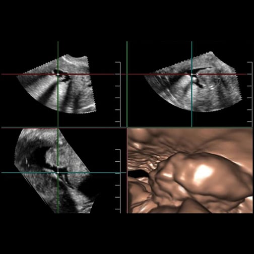
The Aplio 500's Fly Thru technology allows exploration of fluid-filled interior ducts and vessels during or after an examination, and Smart Fusion technology synchronizes ultrasound imaging with CT or MR imaging.
With ever-increasing concerns about radiation dose and the appropriate use of diagnostic imaging tests, doctors are continuously looking for ways to better image their patients. Advances in ultrasound are making the technology appear more attractive for certain clinical applications, from breast health to cardiology, thanks to their noninvasiveness, cost-effectiveness and lack of radiation. Some of the emerging innovations in ultrasound, such as real-time 3-D imaging and the development of wireless transducers, are set to keep the market going throughout 2013 and beyond.
More Mobility
One of ultrasound’s key advantages over other imaging modalities is its increasing mobility. From innovations such as portable hand-held devices to the world’s first wireless transducer, ultrasound has the ability to quickly image internal organs — such as the heart, kidney and liver — without worrying about troublesome cables or moving bulky machines around in a patient’s room. This key feature of ultrasound can improve not just the point of care at hospitals, but also the public’s access to medical imaging, especially in developing regions where it is needed.
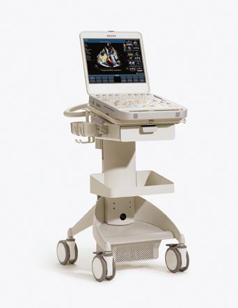
Companies are also working to improve compact ultrasound systems for use in tight premium spaces within hospitals, such as in the operating room. At the 2012 Radiological Society of North America’s (RSNA) annual meeting, Philips announced it now offers transducers for surgical applications on its compact CX50 ultrasound systems.
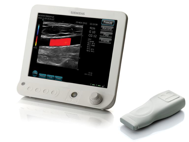
Also at RSNA 2012, Siemens Healthcare introduced the world’s first ultrasound system with wireless transducers, the Acuson Freestyle. Completely untethered from the console, the Acuson’s wireless transducer can be used to image from up to 10 feet away, and is built to endure any potential falls or mishaps, according to Michael Cannon, vice president and general manager of point-of-care solutions at Siemens Healthcare. The system is targeted toward use in interventional applications such as radiology and cardiology, and Cannon said it could also be used to aid in vascular access procedures as well as needle visualization. The system uses a proprietary 8 GHz ultra-wideband radio frequency to transmit data to the main console, and also includes Bluetooth wireless control.
Enhancements to Traditional Ultrasound
Other recent technological trends in ultrasound include advances for contrast-enhanced imaging, volume imaging and elastography. In addition to providing real-time imaging of internal anatomy, these advances in ultrasound allow doctors to image blood perfusion and blood flow, view real-time 3-D imaging of structures, and more easily differentiate malignant tumors from the benign, among other functions.
The availability of contrast-enhancing microbubbles gives doctors the ability to more clearly delineate between tissue and fluids, said Edward Leen, M.D., FRCR, professor of radiology at Imperial College London. By enhancing the reflection of ultrasound waves, microbubbles create a clearer contrast between individual organs and blood flow in the image. Contrast agents include Bracco’s SonoVue, widely used in Europe, and Lantheus’ Definity, which is more common in the United States, according to Leen. Other features of contrast-enhanced ultrasound (CEUS) include the ability to display vascularity in rendering modes and to more easily characterize malignant liver lesions for oncology applications. Untargeted CEUS currently is used clinically in applications such as echocardiography, while targeted CEUS remains in development.
Additionally, limitations with 2-D ultrasound for interventional applications have many clinicians looking toward 3-D and 4-D ultrasound for real-time volume imaging, made possible by the continuous increase of computing power in ultrasound systems. RSNA 2012 marked the one-year anniversary of Toshiba’s launch of the Aplio 300 and 500 systems, the latter of which offers 3-D and 4-D applications for clinical use. The Aplio 500’s Fly Thru technology allows operators to “fly through” and explore fluid-filled interior ducts and vessels during or after the examination. Its Smart Fusion technology provides the ability to synchronize ultrasound imaging with computed tomography (CT) or magnetic resonance (MR) image, improving the detection of hard-to-find lesions.
In combination with advanced visualization functions, 3-D ultrasound can aid complex surgical applications and interventional procedures, according to Leen. Images are reconstructed from echoes sent by the transducer from multiple angles, and with 4-D ultrasound real-time moving images can be produced. Multiplanar reconstructed (MPR) images are now available for review in the same manner as CT and MR scans, Leen said.
And in comparison to other modalities, 3-D/4-D ultrasound has a more rapid acquisition rate of datasets and subsequent improved image visualization, thanks to the advent of matrix array transducers. These latest transducers are also becoming smaller in size than previous generations, Leen said. This in turn can improve various parts of the workflow — namely, shortening exam times and reducing the risk of repetitive strain injury (RSI) for operators.
Furthermore, he said 3-D advances are making way for functional imaging with ultrasound, as manufacturers start integrating contrast-enhanced capabilities into their new systems. The overall lower costs of high-end ultrasound systems in comparison to other modalities also add to their cost-effectiveness and appeal to many hospitals.
Innovations in elastography are also making waves in the ultrasound market. Providing the capability to differentiate between soft normal tissue and stiffer malignant tumors, color-coded elastography maps can be produced by several of the latest ultrasound systems. One technological advance that continues to produce clinical results since its approval by the U.S. Food and Drug Administration (FDA) in 2009 is ShearWave Elastography (SWE), developed by Supersonic Imagine. SWE differs from traditional strain elastography, which relies on manual compressions by the examiner to assess tissue displacement, according to Peter N. Burns, Ph.D., a professor at the University of Toronto. The SWE technique instead captures shear wave generation with patented UltraFast Imaging technology — currently only available on the Aixplorer ultrasound system from Supersonic — to quantitatively measure the stiffness of local tissues. By being less dependent on an experienced operator, SWE can record elasticity more accurately than conventional elastography and produce a more objective, real-time color-coded map to show tissue stiffness.
In October 2012, Supersonic announced FDA clearance of a real-time adjustable numerical scale (ANS) in meters per second for the technology, allowing clinicians to adjust the scale to optimize visualization of elasticity according to the organ of interest. This adds to the appeal of SWE, since stiffer tissues can vary among different pathologies. Currently it is widely used and studied in breast health applications. In March 2012, results from a multicenter study, published in the Radiology and European Radiology journals, showed the technology improves breast ultrasound specificity when detecting breast lesions in patients.
Innovation Continues
While traditional 2-D ultrasound continues to be widely adopted by a broad range of clinical applications, recent technological advances are opening more areas of interest in the 3-D and 4-D ultrasound markets. Software improvements and more sophisticated computing algorithms are allowing manufacturers to offer smaller, more powerful and more complex systems. These innovations will continue to expand as physicians compare ultrasound with other imaging modalities when choosing how to best diagnose their patients and plan interventional procedures. itn

 November 03, 2025
  clearance")