Multiple studies and products were presented at the 2014 Radiological Society of North America (RSNA) conference in December about emerging technologies in breast imaging, with a focus on how they will affect women who have dense breasts. Most researchers have been comparing the utility of mammography screening versus tomosynthesis.
According to Volpara Solutions, for a significant number of women, the efficacy of mammography is reduced due to the masking of cancers in areas of increased tissue density. Ultrasound and magnetic resonance imaging (MRI) are two supplementary breast imaging modalities that retain their sensitivity in women with dense breasts, and when used in addition to mammography, can demonstrate an increased cancer detection rate compared to mammography alone.1
However, both modalities have higher rates of false-positive findings, which can often result in more tests and unnecessary biopsies, making MRI and ultrasound expensive to implement in high-volume screening programs, according to the study “3-D Mammography Improves Cancer Detection in Dense Breasts” presented at RSNA 2014.
A newer breast imaging modality, digital breast tomosynthesis (DBT) or 3-D mammography, creates a 3-D picture of the breast using X-ray,2 and may become a more popular method of screening. According to researchers, using a tomosynthesis dataset greatly reduces detection challenges associated with overlapping structures in the breast, which is the primary drawback of conventional 2-D analog and digital mammography.
According to the study, “Tomosynthesis in Breast Cancer Visualization as a Function of Mammographic Density,”3 3-D mammography was beneficial for visualizing noncalcified breast cancers in scattered and heterogeneously dense breasts with about 70 percent of cancers in these density categories seen only with tomosynthesis. Patients with fatty and extremely dense breasts had cancers seen equally well with tomosynthesis and 2-D mammography.
Hologic conducted a multi-center clinical trial that compared 2-D digital mammography plus tomosynthesis imaging (combo mode) to 2-D mammography alone. Results found that the addition of tomosynthesis to digital mammography increased diagnostic accuracy and reduced recall rates for non-cancer cases.
The addition of 3-D imaging has been shown to improve the performance of mammography in both fatty and dense breasts, according to Hologic. Because dense breasts have more fibroglandular tissue than fatty breasts, it was expected that 3-D mammography would provide improved performance in denser breasts. However, clinical data shows that 3-D mammography was helpful for all breast densities.
Emerging Mammography Modalities
Hologic 3-D mammography is a combined examination of 3-D and 2-D imaging. It takes only seconds longer than conventional 2-D digital mammography, and can assist in increased cancer detection (by 27 percent), increased invasive cancer detection (by 40 percent) and decreased callback rates (20-40 percent), localizing structures in the breast and improved lesion and margin visibility.4
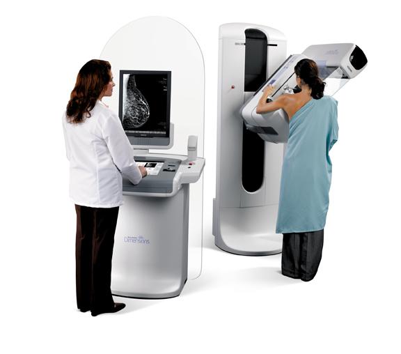
Hologic’s Selenia Dimensions mammography system offers multiple workstations, including a flexible platform that allows users to start with both 2-D digital mammography and Hologic 3-D mammography for screening, diagnostic and interventional procedures, or with a 2-D digital mammography-only system; controls featuring icons and function screens plus a system that provides scans that take less than four seconds, a 15-degree scan that provides high in-plane resolution and the acquisition of 15 projection images (one image/degree of arc) and filtered back projection.5
The optimized face shield is retractable to help with positioning patients and is stationary during 3-D imaging allowing for patients to be positioned as they are for 2-D imaging. The Fast Paddle system conforms to the natural contours of the breast, which provides for better comfort to the patient and even more compression across the breast. All paddles are used for 2-D and 3-D imaging.
Designed for use with the Selenia Dimensions mammography system, Hologic’s MammoPad cushion provides a comfortable surface between the patient and the image receptor, helping the patient relax the pectoral muscle.6 This allows the technologist to better position the breast for screening and to obtain more of the chest wall image. The cushion’s grip-like surface holds the breast tissue in place, making the patient more relaxed, which helps the technologist position them correctly, saving time and improving image accuracy.
Radiation dose management for breast imaging has been on researchers’ radar as healthcare providers search for ways to reduce the amount of radiation patients are receiving if they need to have supplementary screening. Hologic’s U.S. Food and Drug Administration (FDA) approved C-View software option generates images that can be used in place of traditional 2-D images as part of the 3-D breast exam. The 2-D and 3-D image slices are reviewed together. The software makes lower dose exams possible, and the combined 3-D and C-View exam offer better performance compared with just 2-D.7
Molecular Breast Imaging
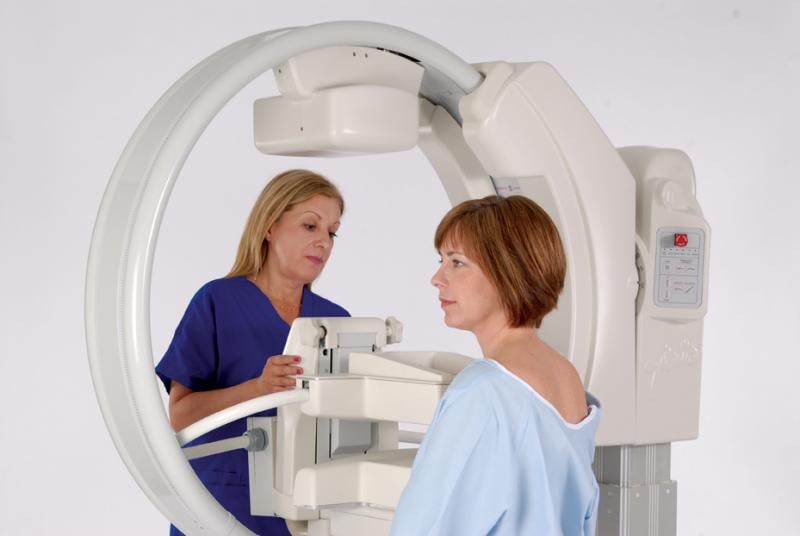
Molecular breast imaging (MBI) is a highly effective secondary diagnostic tool, particularly for women with dense breast tissue.8 Gamma Medica’s LumaGEM MBI technology measures and images the distribution of radionuclides by means of photon detection in order to aid in the evaluation of lesions in the breast tissue.9 The product’s dual-head technology identifies tumors in dense breast tissue where findings are inconclusive or even negative with film-based and digital mammography, and is able to detect breast cancers as small as 5 mm that may have been missed by mammography alone.
Updates in Radiology Reading Rooms/Displays
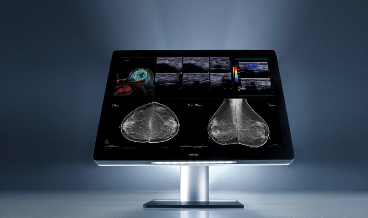
Radiology reading rooms are seeing a facelift with new display systems that address ergonomics and multi-viewing modalities. The Coronis Uniti product from Barco, which combines PACS and breast images on one screen, was created in response to specific challenges that are occurring in modern radiology: increasing image volumes, growing complexity and occupational discomfort.10 The 12 MP screen offers images in calibrated colors and gray scales while allowing the viewing of 2-D, 3-D, static and dynamic images such as computed tomography (CT) moving images, ultrasound, magnetic resonance imaging/positron emission tomography (MRI/PET) scans, multi-frame mammography and digital breast tomosynthesis. With the ability to view multi-modality images, radiologists no longer have to travel to separate reading rooms, therefore increasing workflow productivity.
To ease physical discomfort when reading images, Barco designed a display to mirror a human’s natural field of vision and has a 33-inch screen size and Fusion form factor to create a canvas for side-by-side comparison of multiple high-res images. This format minimizes head and eye movement and reduces the need for scaling and rearranging to complete studies more efficiently. To reduce eye fatigue, the SoftGlow wall light adds ambient light to the reading room while the SoftGlow task light shines light on papers and film folders. Barco’s Optical Glass technology is incorporated into the display, which reduces reflections and enhances image sharpness.
Barco’s RapidFrame technology allows radiologists to view moving images in-focus and features a built-in light box for viewing of 18 x 24 or 24 x 30 films. This allows for the comparing of current digital exams with film-based priors without leaving the workstation.
The Mammo Tomosynthesis 5 MP from Barco is a display system available for breast tomosynthesis. Cleared by the FDA, the display system is used for standard and multi-frame digital mammography as well as breast tomosynthesis. The Mammo Tomosynthesis 5 MP instantly delivers images without motion blur.11 The display system includes RapidFrame, per pixel uniformity (PPU measures and adjusts the luminance of each pixel, making them DICOM compliant), SmoothGray (generates grayscale images), DuraLight Nova (backlight of 1,000 cd/m²), I-Luminate (button will temporarily boost display brightness), I-Guard (sensor continually guards and adjusts luminance output), QAWeb (Web-based tool for automated calibration) and an adjustable dual-head stand, which allows users to angle the displays.12
With technology and research continuing to prove that tomosynthesis may have the upper hand in early detection of breast cancer, and allowing for better screening results for women with dense breasts, healthcare providers may make traditional mammography a method for secondary screening instead of a first-choice method. itn
References:
1. www.volparasolutions.com/solutions/volparadensity/, accessed Jan. 27, 2015.
2. www.breastcancer.org/symptoms/testing/types/dig_tomosynth, accessed Jan. 28, 2015.
3. Philpotts LE, Raghu M, Geisel JL, et al. “Tomosynthesis in Breast Cancer Visualization as a Function of Mammographic Density.” Radiological Society of North America annual meeting, Chicago, 2013.
4. www.hologic.com/sites/default/files/DS-00194_SeleniaD_5000_US_Tomo_Rev%20002_11-13.pdf, accessed Jan. 28, 2015.
5. www.hologic.com/products/imaging/mammography/selenia-dimensions-mammography-system, accessed Jan. 28, 2015.
6. www.hologic.com/products/imaging/mammography/mammopad-breast-cushion, accessed Jan. 28, 2015.
7. http://investors.hologic.com/2013-05-21-Hologic-Receives-FDA-Approval-for-a-New-Low-dose-3D-Mammography-Breast-Tomosynthesis-Solution-for-Breast-Cancer-Screening, accessed Jan. 28, 2015.
8. www.gammamedica.com/women-and-molecular-breast-imaging/faq/, accessed Jan. 28, 2015.
9. www.gammamedica.com/our-products/lumagem/, accessed Jan. 28, 2015.
10. www.barco.com/en/uniti, accessed Jan. 28, 2015.
11. www.barco.com/en/Products-Solutions/Displays-monitors-workstations/Medical-displays/Mammography-displays/5-MegaPixel-display-system-for-digital-breast-imaging-including-breast-tomosynthesis.aspx, accessed Jan. 28, 2015.
12. www.barco.com/en/Products-Solutions/Displays-monitors-workstations/Medical-displays/Mammography-displays/5-MegaPixel-display-system-for-digital-breast-imaging-including-breast-tomosynthesis.aspx?tab=features, accessed Jan. 28, 2015.

 April 24, 2024


