
Greg Freiherr has reported on developments in radiology since 1983. He runs the consulting service, The Freiherr Group.
Blog | October 20, 2011
Toward a Better, Gentler Way to Image Breast Cancer
Minimizing X-ray dose has driven the global imaging community to develop technologies that today can cut patient dose from computed tomography (CT) to a fraction of its previous levels. So it may soon be in mammography.
Earlier this year, the U.S. Food & Drug Administration (FDA) granted permission for Hologic to sell its Selenia Dimensions, a machine that through its ability to perform tomosynthesis offers mammographers a chance to see what otherwise might be hidden. Multiple X-ray exposures at varying angles promise to expose lesions obscured by overlying tissues and increase diagnostic certainty.
Unfortunately, to achieve the full potential of this technology, the tomo scan must be read in the context of a 2-D mammogram. This requires two exams, doubling the patient’s dose of radiation. While little has been said so far about this, just as it was for CT, dose could become an issue for tomosynthesis, giving pause to some whose patients might otherwise benefit from tomo. But work is already underway to create an arsenal of silver bullets.
Researchers at the University of Pittsburgh hope to craft one such a bullet against dose, an algorithmic one in the shape of a “synthetic full-field digital mammography (FFDM)." The goal is to synthesize data for a 2-D image from those obtained during the tomo scan, thus sparing the patient from a second exposure to radiation. If successful, the tomo exam would capture all the data mammographers need.
Promising to improve tomo further is a nanotechnology that could increase scan speed by a factor of 10, boosting spatial resolution and opening the door to dual-energy scanning, potentially providing better contrast and, in the process, reducing radiation dose. This technology would replace the moving X-ray source in tomo with a stationary one, called multi-pixel field emission X-ray, which is based on microscopic carbon nanotubes. The feasibility of doing so has already been demonstrated and a fully functional system is in development at a company called XinRay Systems, a joint venture between Siemens and a small North Carolina startup called Xintek.
On the detector side, at the State University of New York in Stony Brook, researchers are working on a new amorphous selenium detector that could double the detective quantum efficiency (a measure of X-ray detector performance), boosting resolution far beyond what is currently possible and, potentially reducing dose.
With its volumetric data sets providing different angles on suspicious lesions, today’s breast tomosynthesis is hard to argue against. Tomorrow it may be even more so, as the evolution of this technology renders issues about dose less and less of an issue.
Related Content

News | Artificial Intelligence
May 15, 2024 — Heart disease is the leading cause of mortality in the U.S., accounting for one out of every five deaths ...
 May 15, 2024
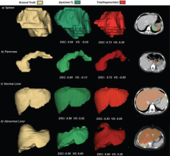
News | Pediatric Imaging
May 15, 2024 — Transfer learning (TL) models trained on heterogeneous public datasets and fine-tuned using institutional ...
May 15, 2024 
News | Radiology Business
May 14, 2024 — University Hospitals (UH) and Siemens Healthineers announce a 10-year strategic alliance that builds on ...
May 14, 2024 
News | Breast Imaging
May 10, 2024 — According to the Summa Cum Laude Award-Winning Online Poster presented during the 124th ARRS Annual ...
May 10, 2024 
News | Treatment Planning
May 6, 2024 — Elekta announced the acquisition of Philips Healthcare’s Pinnacle Treatment Planning System (TPS) patent ...
May 06, 2024 
News | Mammography
May 6, 2024 — Enable Me, a VELA Medical company, cited major new research by Siemens Healthineers entitled, “The future ...
May 06, 2024 
Feature | Radiology Business | By Melinda Taschetta-Millane
One on One interviews with radiology trailblazers and historic FDA clearances made the top-read list for April. Take a ...
May 03, 2024 
Feature | Breast Density | By Robert L. Bard, MD
Decades since the advent of breast scanning technology, innovations in noninvasive diagnostic imaging provide new ...
May 03, 2024 
News | Pediatric Imaging
May 2, 2024 — Head and abdominal trauma is a leading cause of death for children. About 1%–2% of children who come to ...
May 02, 2024 
Feature | Radiology Business
Beginning this spring, ITN will begin sending out a bi-monthly survey to our readers on a variety of topics, which we ...
May 02, 2024 © Copyright Wainscot Media. All Rights Reserved.
Subscribe Now