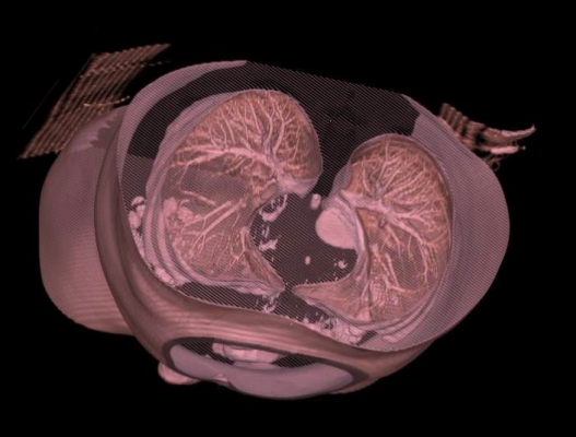
August 28, 2013 — Medical software developer Blackford Analysis announced that picture archiving and communications systems (PACS) integrated with its automatic alignment technology deliver significant time savings when matching lung nodule locations across current and prior chest computed tomography (CT) exams. Preliminary results indicate a time saving of greater than 50 percent with automatic deformable alignment over manual alignment of current and priors.
Lung nodules, a common finding in chest CT exams, are routinely followed by serial chest CT where the radiologist must assess individual nodules for change. Manually matching nodule location across exams is burdensome due to differences in pulmonary anatomy caused by variance in patient positioning and breath hold.
The Stony Brook study, which will be published later this year, retrospectively identified 27 subjects from Stony Brook Medical Center with nodules in two chest CTs within three years. The study measured the time taken for board-certified radiologists to match each of the 112 nodules in prior exams to the same location in current exams in PACS using two volume alignment methods:
- Manual Rigid, where the radiologist synchronized scrolling manually at the most superior slice where air was visible in the right lung.
- Automatic Deformable, where Blackford Analysis’ PACS-integrated software performed automatic deformable alignment of current and prior chest CT exams.
Preliminary results show that automatic deformable alignment provided by Blackford Analysis significantly reduces lung nodule location matching time when compared with conventional manual slice synchronization. Preliminary data has shown that automatic deformable alignment can reduce location match time by greater than 55 percent compared to manual alignment.
“Lung nodule studies can be quite a challenge to compare due to the differences in pulmonary anatomy caused by variance in patient positioning and breath hold,” said Matthew A. Barish, M.D., FACR, clinical associate professor of radiology; director, body imaging; director, body MRI (magnetic resonance imaging); Stony Brook Medicine. “Using a PACS integrated with Blackford Analysis has made a clear difference to our ability to quickly identify nodule locations across exams, and our early data confirms a significant reduction in the time spent locating nodules in follow-up studies allowing for quick determinations of nodule stability or growth.”
The U.S. Preventive Services Task Force (USPSTF) recently issued a draft recommendation in favor of low-dose CT lung cancer screening for individuals aged 55-79 with at least a 30 pack year smoking history who currently smoke or quit within 15 years.
“This data is particularly relevant in light of recent developments, as the USPSTF recommendation on lung cancer screening is going to have a big impact on radiologists across the United States,” said Ben Panter, CEO, Blackford Analysis. “Once a reimbursement code is issued, there will be a massive increase in low-dose chest CT screening studies for lung cancer, with an increasing number of priors to compare for change in lung nodules, which will present a significant increase in workload for radiologists, and that’s where our software can really help make a positive difference.”
Designed to be seamlessly integrated into any PACS, the Blackford software suite uses algorithms to allow rapid alignment of current and prior cross-sectional exams, including those from different modalities such as CT, MRI and PET (positron emission tomography).
For more information: www.blackfordanalysis.com


 February 09, 2026


