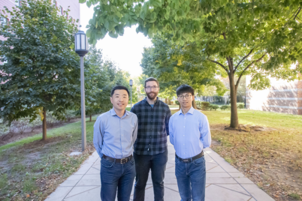
(From left): Pengfei Song, a researcher at the Beckman Institute and a professor of electrical and computer engineering and bioengineering; Matthew Lowerison, a Beckman Institute Postdoctoral Fellow; and Zhijie Dong, a Ph.D. student in the Song Lab, are members of an interdisciplinary, multi-institute team that received NIH funding to develop a new device that can instantly add 3D capabilities to 2D ultrasound imaging systems. Their affordable, user-friendly design could help make high-quality medical imaging more accessible in diverse communities. Beckman Institute for Advanced Science and Technology.
October 13, 2022 — A new device in the works at the Beckman Institute for Advanced Science and Technology could make high-quality medical imaging more accessible in diverse communities. Affordable and user-friendly, it is designed to instantly add 3D capabilities to 2D ultrasound imaging systems.
Pengfei Song, a researcher at the Beckman Institute and an assistant professor of electrical and computer engineering and bioengineering at the University of Illinois Urbana-Champaign, is leading the project, which is supported by a four-year, $2M award from the National Institute of Biomedical Imaging and Bioengineering at the National Institutes of Health.
“Ultrasound is one of the most cost-effective medical imaging technologies,” Song said. “It’s the natural first stop in increasing accessibility to many clinical applications, such as providing care for patients suffering from cardiovascular diseases or cancer.”
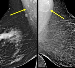
Like bats slaloming around stalactites, guided by echolocation, humans use ultrasonic waves to visualize the body’s internal landscape. Ultrasound imaging typically uses a handheld probe to send a beam of ultrasonic waves toward a target, like a tumor; clinicians can determine its size, shape, and location based on how the waves bounce back. Cancer care providers use ultrasound to identify abnormal tissues and tumors for screening and diagnosis.
But unlike the nocturnal echolocators of the animal kingdom, clinical ultrasound systems mostly operate in 2D, which restricts the range of view during a scan.
“The human anatomy is three-dimensional,” Song said. “A 2D ultrasound image of a tumor, tissue, or internal organ can be difficult to interpret.”
During a 2D ultrasound scan, a slight change in the angle of the probe or the patient’s posture can make objects appear larger or smaller than they are.
“With 3D ultrasound, you can capture the whole object and surrounding environment, and you have landmarks to know exactly what you’re looking at,” said Matthew Lowerison, a Beckman Institute Postdoctoral Fellow in the Song Lab.
The researchers’ proposed device uses a clip-on technique to easily integrate with the 2D ultrasound probes that most clinics already own. 3D systems are available in some clinics, but they are mainly used in high-end facilities for specialized care.
“Clinicians have different probes for imaging different body parts, and our device can clip on to the vast majority of those,” Lowerison said.
Aptly named FASTER, the device is designed to instantly enable real-time 3D ultrasound imaging for clinics in diverse communities, especially those where 3D imaging is cost prohibitive.
“We want 3D ultrasound imaging to be a possibility wherever 2D ultrasound imaging is used,” said Zhijie Dong, a Ph.D. student in the Song Lab who is leading the technology development for FASTER. “We hope that access to 3D technology will drastically improve the level of care that clinics can offer.”
In FASTER’s first clinical application, which will be conducted in collaboration with the Mayo Clinic in Rochester, Minnesota, the researchers will focus on imaging axillary lymph nodes on patients with breast cancer.
In FASTER’s first clinical application, which will be conducted in collaboration with the Mayo Clinic in Rochester, Minnesota, the researchers will focus on imaging axillary lymph nodes on patients with breast cancer.
“As a practicing radiologist with a specialty in breast imaging, I am very excited to be part of this innovative research proposal. I can immediately see the clinical usefulness of this new technology for improving the performance of breast ultrasound,” said Dr. Katrina Glazebrook, a radiologist at the Mayo Clinic who will collaborate on this project alongside colleague Dr. Shigao Chen.
The versatile model means that as imaging technology evolves, FASTER will, too.
“With the rapid development of pocket-sized, handheld ultrasound imaging systems, more and more ultrasound imaging procedures can be done by individuals without formal sonography training. 3D imaging is essential in these situations because non-experts can scan the general location in need of attention, and a physician can interpret the images,” Song said.
While a 2D ultrasound scan uses a stationary probe to direct a beam of sound waves in a fixed direction, a 3D ultrasound scan sweeps the beam back and forth. The two most common methods of doing this — manual rotation or automatic motors — can be unwieldy and impractical for scaled-up use.
“We needed a compact, fast, and accurate solution to move the ultrasound beam,” Song said.
With small, fast-tilting mirrors, FASTER can sweep the ultrasonic waves while the probe itself remains stationary, a key innovation that aims to make 3D imaging systems faster and more compact.
“It’s all about thinking creatively,” Song said. “If you can’t move the source, move the signal.”
In addition to clinical applications, FASTER could significantly impact basic research in ultrasound, as the device provides a robust, low-cost solution for ultrafast 3D imaging, the foundation for many advanced ultrasound imaging methods, such as shear wave elastography, functional neural imaging, and super-resolution imaging.
For more information: https://beckman.illinois.edu/

 May 17, 2024