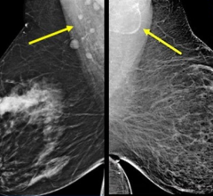
\"In retrospect, we might have seen the larger, 8 mm cancer on the 2D images,\" explains Marcela Bohm-Velez, M.D., FACR, \"But the second cancer was extraordinarily subtle -- only 3 mm in size --and there is no way we would have seen it on the 2D images.\"
Just weeks after implementing 3D mammography (breast tomosynthesis) in their practice, radiologists at Weinstein Imaging Associates found breast cancers in three patients that were not seen on the 2D images and the entire staff became instant believers in the new technology.
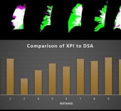
In one patient, the 3D images identified two cancers. “In retrospect, we might have seen the larger, 8 mm cancer on the 2D images,” explains Marcela Böhm-Vélez, M.D., F.A.C.R., President of Weinstein Imaging. “But the second cancer was extraordinarily subtle — only 3 mm in size — and there is no way we would have seen it on the 2D images.” In two other patients, the first cancers were visible on the 2D images, but the 3D images showed second, smaller cancers that were not seen on 2D.
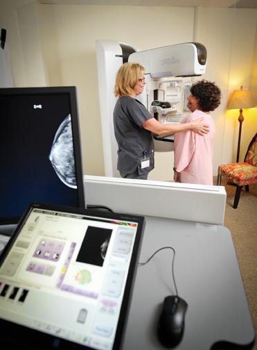
Weinstein Imaging Associates is a premier breast imaging center in the Pittsburgh area with three offices providing a full suite of women’s imaging services, including mammography, breast MRI and stereotactic and ultrasound-guided biopsies. “If we see something suspicious on a mammogram, we work it up the same day, often doing additional imaging, ultrasound or a biopsy, if needed,” explains radiologist and Vice President Michelle R. Straka, M.D. “We don’t want to make patients come back for multiple visits or suffer through the anxiety of waiting and wondering whether they have breast cancer.”
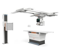
Böhm-Vélez followed the development of 3D mammography closely and knew she wanted the technology for her patients when it became available. As soon as it was approved, Weinstein Imaging added a Hologic Selenia Dimensions breast tomosynthesis system to one of its three offices in the Pittsburgh, Pennsylvania, area.
“3D mammography supports our commitment to provide patients with the latest, most innovative technology for breast cancer detection,” states Böhm-Vélez. “It allows us to detect cancers earlier and reduce the need for additional imaging.”
Performance in Dense Breasts Reduces Need for Ultrasound
Weinstein Imaging has found 3D mammography to be particularly effective for women with dense breast tissue, providing a considerable advantage to patients and the radiologists interpreting the images. According to Böhm-Vélez, “The sensitivity of imaging is low when dense breast tissue is involved, making it difficult to identify abnormalities. 3D mammography separates the breast tissue allowing us not only to see the abnormality better, but also to determine whether it is a mass, or just overlapping tissue.” Straka adds, “For some patients with dense breast tissue, the images uncover a previously undetectable nodule or mass.”
Böhm-Vélez continues, “Fifty percent of women have dense breasts and it can be quite a challenge to find small cancers in these mammograms. 3D mammography increases our confidence that there is not a small tumor hiding between the breast tissues. Our objective has always been to detect early stage cancers since it has been shown that breast cancer is a treatable disease.”
Fewer False Positives, Fewer Unnecessary Tests
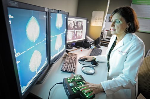
Another key benefit of mammography is the ability to reduce false positives. After screening over 900 patients using the Hologic system, Weinstein Imaging has seen a substantial reduction in the need for additional views and ultrasounds in patients where overlapping tissue was the question. “3D mammography lets us scroll through all the breast tissue in 1 mm slices, which takes away tissue overlap and the false positives generated from tissue overlap,” states Straka.
Prior to adopting 3D mammography, each time Weinstein Imaging identified a new breast cancer, they routinely performed an ultrasound, because when new breast cancers are detected, a second cancer will be present in approximately 10 percent of patients. “Now, the 3D images help us identify the second cancer,” says Straka.
More Patients Choose 3D
Weinstein Imaging offers 3D mammography to every patient, screening and diagnostic, and, in the few months since they began, more than 30 percent of women have opted to have the 3D exam. “We provide patients with the innovative technology and accurate interpretation they would find in leading hospitals, but in the environment of a private practice,” explains Straka. “I think 3D mammography is bringing new patients to our practice.” In fact, as a result of the practice’s early educational efforts, many referring physicians are now coming to Weinstein Imaging for their own mammograms.
“Our mission is to provide high-quality, innovative imaging and personalized patient care,” concludes Böhm-Vélez. “3D mammography is beneficial to my patients and that is what I am always looking for.”
Case study supplied by Hologic Inc.
The views and opinions expressed herein are those of Marcela Böhm-Vélez, M.D., F.A.C.R., and her colleagues at Weinstein Imaging and are not necessarily those of Hologic.
This information is intended for medical professionals in the U.S. and other markets and is not intended as a product solicitation or promotion where such activities are prohibited. Because Hologic materials are distributed through websites, eBroadcasts and trade shows, it is not always possible to control where such materials appear. For specific information on what products are available for sale in a particular country, please contact your local Hologic representative or write to [email protected].
Hologic, Dimensions and Selenia are trademarks and/or registered trademarks of Hologic and/or its subsidiaries in the U.S. and/or other countries.
 May 17, 2024