The most common cause of chronic liver disease? Nonalcoholic fatty liver disease (NAFLD). With 25% of the world’s population diagnosed, NAFLD is the most common cause of chronic liver disease globally.1,2 Of patients diagnosed with NAFLD, approximately 20-30 percent progress to more advanced NASH fibrosis.3
Fatty liver, or hepatic steatosis, occurs when fat molecules are not metabolized efficiently enough by the body and end up stored in the liver. While a normal liver contains a small amount of fat, the liver is considered “fatty” if the amount of fat within the hepatocytes exceeds 5 percent.4
The liver’s primary role is to filter blood and remove harmful substances. When the liver detects something harmful, it works to eliminate and clear that substance from the body. During this process and immune response, some inflammation occurs. Yet, the liver is a fascinating organ. Once that harmful substance is cleared, the liver inflammation resolves, and the liver is once again healthy.
The problem comes when there is a constant inflammatory response, which could stem from a variety of behaviors or causes. As inflammation continues, liver tissue becomes increasingly stiff, and fibrosis may develop. If left untreated, the liver tissue may be unable to repair itself. This causes permanent and irreversible liver damage called cirrhosis.
Early detection of hepatic steatosis improves the chances of managing or reversing the condition before changes that require expensive medical intervention or irreversible changes occur.2
The Real Cost and Availability of Fatty Liver Diagnosis Techniques
In the United States, the annual direct medical costs for nonalcoholic fatty liver disease are estimated to be about $103 billion.5
There are numerous imaging and non-imaging medical techniques that diagnose hepatic fat content. However, these techniques vary in effectiveness, cost, invasiveness, availability of equipment, and/or patient suitability.
Currently, liver biopsy is the reference standard for diagnosing and grading hepatic steatosis. Liver biopsy is, however, inappropriate for screening or frequent monitoring, as it is invasive, prone to sampling errors, observer variability, and the risk of complications.3
When it comes to characterizing fat content in the liver, MRI-PDFF is the most widely accepted noninvasive imaging modality. MRI-PDFF is a quantitative imaging biomarker that enables accurate, repeatable, and reproducible quantitative assessment of liver fat over the entire liver.3 It provides a specific fat content number, between 1–100 percent. Yet, the cost, exam time, and ease of availability makes routine MRI-PDFF challenging. Due to these challenges, there is a need for technology that is less invasive, more cost-effective, and widely available. Here, the advantages of using ultrasound to help diagnose hepatic fat content become clear.
An Accurate AND Accessible Dagnosis Tool
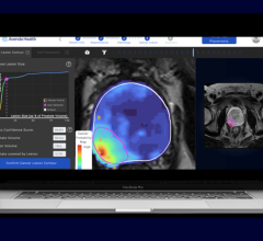
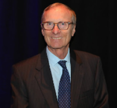
“With the association between hepatic steatosis [fatty liver] and fibrosis progression, it is critical to have accurate, noninvasive tools to aid in not just assessing fibrosis but also hepatic steatosis,” said Richard G. Barr, M.D., Ph.D., at Southwood’s Imaging. “With the ACUSON Sequoia ultrasound system, there are two new quantitative tools to meet this need: Auto Point Shear Wave Elastography [Auto pSWE] and an Ultrasound Derived Fat Fraction [UDFF] index. Auto pSWE enables up to 15 valid pSWE measurements literally in seconds; along with it comes a UDFF index that like the MRI Proton Density Fat Fraction (PDFF) classifies hepatic steatosis as an index value greater than 5 percent.”
A radiologist for more than 25 years, Barr is president of Radiology Consultants, Inc., and a diagnostic radiology specialist at Southwood’s Imaging, both in Youngstown, Ohio. He believes the ability to quantitatively detect hepatic steatosis in the early stages of chronic liver disease would be invaluable for referring physicians. “This could allow them to treat patients with hepatic steatosis earlier, monitor their progression, and work to minimize the incidence of advanced progression through early treatment and lifestyle changes,” he said.
.png)
Expanding Ultrasound’s Role in Liver Assessment
Ultrasound has many obvious advantages, such as reducing ionizing radiation, widespread availability, and improved cost effectiveness over other imaging modalities. But ultrasound also offers other benefits when it comes to liver assessment since it already plays a critical role in the diagnosis and management of liver disease. In fact, ultrasound is currently used to examine liver size, texture, vascularity, and liver tissue stiffness, as well as the identification and characterization of focal lesions.
A key quantitative tool to aid in the diagnosis and treatment of liver fibrosis is liver elastography, which measures shear waves in the liver that are proportional to liver tissue stiffness. The higher the shear velocity, the stiffer the tissue. As a result, liver elastography is a crucial tool in liver fibrosis diagnosis.
The current methods of assessing fatty liver using conventional ultrasound are qualitative. Radiologists will often grade the degree of fatty infiltration based on a variety of characteristics in the B-mode image, which can be subjective. There would be great clinical value in a quantitative ultrasound method to identify hepatic steatosis (fatty liver) with a unit of measure and cut off value consistent to that of MR-PDFF.
A Novel Approach
Auto Point Shear Wave Elastography (Auto pSWE) with Ultrasound derived fat fraction (UDFF) are innovative Advanced Liver Analysis tools, available for use on the ACUSON Sequoia ultrasound system.
Liver Elastography is commonly performed in conjunction with a standard ultrasound exam and used as a tool to aid in the diagnosis and treatment of chronic liver disease patients. The liver elastography exam currently requires a repeatable technique since current guidelines require multiple independent acquisitions (5-10), which are performed manually and typically add an additional five minutes to the standard abdominal ultrasound exam. Here, technology that can help reduce liver elastography variability as well as acquisition time can be beneficial for clinicians and patients. Auto pSWE is a new elastography technique on the ACUSON Sequoia designed to address long acquisition times by delivering up to 15 valid pSWE measurements in a single acquisition and in less than 5 seconds. Auto pSWE is faster than conventional pSWE and is as effective as manual measurements.
UDFF can quantify the amount of fat contained within a patient’s liver. Designed to provide a quantitative tool that can be widely accepted and understood, UDFF on the ACUSON Sequoia provides a fat fraction index in a percentage (%) and uses MRI-PDFF as the reference in detecting the presence of steatosis. Therefore, UDFF on the ACUSON Sequoia delivers a similar clinical utility to MRI-PDFF for determining hepatic steatosis. MRI-PDFF and UDFF methods classify hepatic steatosis as an index value greater than 5 percent.2 With the use of this simple tool, clinicians now have a new, widely available, noninvasive measurement to aid in the management and overall assessment of hepatic steatosis. Not only can UDFF be performed in conjunction with a routine abdominal ultrasound, but UDFF can also quantify the amount of fat contained within a patient’s liver in just seconds.6
The Advanced Liver Analysis on the ACUSON Sequoia provides quantitative assessment of liver tissue stiffness and detection of hepatic steatosis in the same acquisition.
Conclusion
As precision medicine expands and the delivery of healthcare evolves, there is a need for quantitative imaging biomarkers that are noninvasive, cost effective, and widely available. The earlier clinicians can detect fatty liver disease—the leading cause of chronic liver disease worldwide—the better the chances of managing or reversing the condition before expensive medical intervention becomes necessary or irreversible changes occur.2 The Advanced Quantitative Liver Analysis for use on the ACUSON Sequoia ultrasound system enables quantitative assessment of both liver stiffness and hepatic steatosis in seconds, addressing many of the current challenges on conventional ultrasound technology. “I find that on the ACUSON Sequoia, I have all of the tools I need for a comprehensive liver assessment, especially on the high BMI patient,” said Barr.
Case study supplied by Siemens Healthineers.
Bibliography
- Younossi Z, Tacke F, Arrese M, Chander Sharma B, Mostafa I, Bugianesi E, Wai-Sun Wong V, Yilmaz Y, George J, Fan J, Vos MB. Global Perspectives on Nonalcoholic Fatty Liver Disease and Nonalcoholic Steatohepatitis. Hepatology. 2019 Jun;69(6):2672-2682. doi: 10.1002/hep.30251. PMID: 30179269.
- Labyed & Milkowski 2021; JUM 39(12) p2427-2428, doi: 10.1002/jum.15364
- Caussy Cyrielle, Reeder Scott B, Sirlin Claude B, Loomba Rohit.
Non-invasive, quantitative assessment of liver fat by MRI-PDFF as an endpoint in NASH trials. Hepatology. 2018 Aug; 68(2):763-772. Doi:10.1002/hep. 29797. - Wilkins T, Tadkod A, Hepburn I, Schade RR. Nonalcoholic Fatty Liver Disease: Diagnosis and Management. American Family Physician. July 1, 2013. Vol 88, Num 1: 35 – 42.
- Clinical Liver Disease Volume 1, Issue 1, pages 2-5, 6 MAR 2012 DOI: 10.1002/cld.3 http://onlinelibrary.wiley.com/doi/10.1002/cld.3/full#fig
- Based on 5 UDFF acquisitions when used as a stand-alone feature.
 May 17, 2024