itnTV

VIDEO: One on One with Amy K. Patel, MD, American Association for Women in Radiology Immediate Past President

Breast Imaging | April 15, 2024

Don't miss ITN's latest "One on One" video interview with AAWR Past President and American College of Radiology (ACR) RAN and RADPAC Chair, Amy K. Patel, MD, discussing advocacy initiatives and innovations in artificial intelligence (AI) for breast imaging

Dr. Patel is a breast imaging trailblazer and radiology advocacy leader. In this video,  learn how radiologists can support key initiatives, ways AI is improving patient care, and more.

Related content:

Leaders from RadEqual and the AAWR Sign MOU, Solidifying Commitment to Advance Opportunities in Radiology

Technology Report: Artificial Intelligence in Radiology 2021

VIDEO: Integrating Artificial Intelligence Into Radiologists Workflow

Recent Video

Enterprise Imaging | September 03, 2021

ITN Editor Dave Fornell collected numerous examples of how PACS and enterprise imaging vendors are improving the speed and workflow of their systems during booth demonstrations at the 2021 Healthcare Information Management Systems Society (HIMSS). The 11 minute video condenses down the highlights of workflow efficiencies seen during two days o vendor booth tours.

There was a clear trend of many vendors moving to new platforms that leverage more modern cloud-platform interfaces. This enables faster study loading speeds over web connections. These platforms are also using deeper integration of third-party applications and artificial intelligence (AI) software that do not require separate logins or workflows. Read more about these key trends observed at HIMSS 2021.

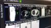
Vendors also showed various ways they have speed up radiology workflows. These included easier to customize hanging protocols, automated fetching of prior exams, synchronizing views and scrolling between a current a prior exams, use of timeline views of patient priors and procedures to make it easier to find relevant images and reports, and integration of all types of images into one unified viewer. 

Specific examples in this video include: 
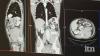
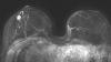
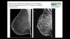
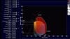
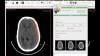
   • Visage Imaging: Example of high speed cloud PACS access to 3D mammograms and and priors. This first video clip shows a demonstration of opening large datasets in a matter of a couple seconds over a network connection from a tethered cellphone.
   • Visage Imaging: Ability to access multiple modalities on one PACS viewer
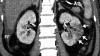
   • GE Healthcare: Examples of fast access to priors and location on screen 
   • GE Healthcare: Example of deep integration of third-party AI software
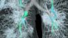
   • Siemens: Overview of its Lung AI Pathway Companion workflow  
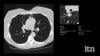
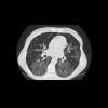
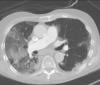
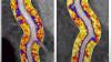
   • Change Healthcare: Enabling fast ability to free rotate around lung anatomy rather than going slice by slice manually 
   • Change Healthcare: Color-coded bar shows loading progress of an image or data set
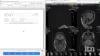
   • Infinitt: Hanging protocol automation to find same view on prior and link for synchronized scrolling   
   • Infinitt: Use of timeline to get quick view of prior reports and images without needing to open whole exam 
   • Siemens: Example of deeper integration with third-party apps, in this case Epsilon strain echo analysis  
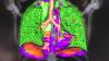
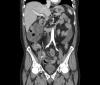
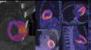
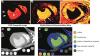
   • Fujifilm: Integrated advanced visualization in the radiology workflow for liver segmentation used for surgical or embolization planning 
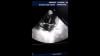
   • Fujifilm: Example of life-like cinematic rendering of a CT scan offers new ways to view anatomy and explain it to a patient 
   • Visage Imaging: Example of enterprise platform able to bring in full original format advanced visualization reconstructed images on a single platform viewer

Related Medical Imaging IT Content From HIMSS 2021:

Advances in CVIS and Enterprise iImaging at HIMSS 21

Photo Gallery of New Technologies at HIMSS 2021

VIDEO: Importance of Body Part Labeling in Enterprise Imaging — Interview with Alex Towbin, M.D.

HIMSS 2021 Showed What to Expect From In-person Healthcare Conferences During the COVID Pandemic

VIDEO: Coordinating Followup for Radiology Incidental Findings — Interview with David Danhauer, M.D.

VIDEO: Cardiology AI Aggregates Patient Data and Enables Interactive Risk Assessments

VIDEO: Examples of COVID-19 CT Scan Analysis Software


Coronavirus (COVID-19) | August 31, 2021

Several radiology IT vendors at 2021 Healthcare Information Management Systems Society (HIMSS) conference demonstrated computed tomography (CT) imaging advanced visualization software software to help automatically identify and quantify COVID-19 pneumonia in the lungs. These tools can help speed assessment of the lung involvement and serial tracking can be used to assess the patient's progress in the hospital and during long-COVID observation. 

Examples of COVID analysis tool shown in this video include clips from booth tours at: 
   • Fujifilm
   • Siemens Healthineers 
   • Canon (Vital)

Canon received FDA clearance for its tool under and emergency use authorization (EUA).

Siemens said its tool was part of its lung analysis originally developed for cancer but modified and prioritized to aid in COVID assessments. 
 

HIMSS Related Content:

Advances in CVIS and Enterprise iImaging at HIMSS 21

Photo Gallery of New Technologies at HIMSS 2021

VIDEO: Importance of Body Part Labeling in Enterprise Imaging — Interview with Alex Towbin, M.D.

VIDEO: Coordinating Followup for Radiology Incidental Findings — Interview with David Danhauer, M.D.

VIDEO: Cardiology AI Aggregates Patient Data and Enables Interactive Risk Assessments

VIDEO: Example of Epsilon Strain Imaging Deep Integration With Siemens CVIS


Information Technology | August 30, 2021

David Danhauer, M.D., FAAP, FHIMSS, chief medical information officer, Owensboro Health, Owensboro, Ky., explains the implementation of healthcare information technology (IT) to coordinate followup on incidental radiology findings. He presented on this topic in a session at the Healthcare Information Management Systems Society (HIMSS) 2021 meeting. 

Their system starts with key words being identified to flag incidental findings by the voice recognition system used to enter radiology report information. IT interfaces with the electronic medical record create a list of patients that need followup and what departments the incidental findings relate to so a coordinator can connect the patient with the proper subspecialty.

Danhauer said many of the incidental findings at his center include lung nodules and abdominal aortic aneurisms. In the past, many of these were lost to followup, but the new system now promotes follow through to get the patient the care they need. This has helped increase revenue, improve patient care and lowers the health system's liability profile. 

The system experienced several patient safety events due to gaps in care coordination with incidental findings documented in the radiology report, but missed by referring physicians. A patient safety initiative he helped implement automating the workflow resulted in a nine-fold increase in identifying and communicating incidental findings for improved patient safety. 

Read about more advances in PACS and enterprise imaging at HIMSS 21.

Photo Gallery of New Technologies at HIMSS 2021

VIDEO: Importance of Body Part Labeling in Enterprise Imaging — Interview with Alex Towbin, M.D. 


Enterprise Imaging | August 27, 2021

Alex Towbin, M.D., Cincinnati Children’s Hospital Medical Center CMIO, Radiology Department associate chief of clinical operations and informatics, and chair of radiology informatics, spoke in an enterprise imaging session at the Healthcare Information Management Systems Society (HIMSS) 2021 meeting and highlight the importance of a standardizing body part labeling to enable imaging consumption, image sharing, greater levels of interoperability and image-based artificial intelligence (AI) research. 

He described the process by which existing body part ontologies were evaluated, how the HIMSS-SIIM Enterprise Imaging Community raised awareness of the issues caused by the lack of an industry-standard body-part ontology, and the process by which an industry standard will be selected. Finally, the speakers will discuss how the HIMSS-SIIM Enterprise Imaging Community plans to advocate for the selected ontology to be incorporated as part of existing standards such as DICOM and HL7 FHIR.

In the video he outlines three metadata elements needed to selection of a relevant comparison imaging examination. He also explains how the HIMSS-SIIM EIC convened experts to select a standard body part ontology for use in enterprise imaging
Describe the HIMSS-SIIM EIC’s plan to foster adoption of a standard body part ontology for use in enterprise imaging
 

Advances in PACS and Cardiology Information Systems at HIMSS 2021

Find more HIMSS content

Enterprise Imaging | August 06, 2021

Integrated Speech recognition solutions are becoming a necessary part of radiology reporting platforms. Konica Minolta recently announced a partnership with nVoq to integrate a speech to text solution into their Exa Platform

ITN recently spoke with Kevin Borden, Vice President of Product, Healthcare IT for Konica Minolta and Chad Hiner, Vice President of Customer Experience for nVoq, to talk about how this integration is improving the Exa user experience.

Related enterprise imaging content:

Talking Trends with Konica Minolta

BLOG: Zero-footprint Viewer with Server-side Rendering Pushes Imaging Forward During Pandemic

BLOG: Exa Gateway Offers a New Way to Deliver Teleradiology 

BLOG: Artificial Intelligence for Clinical Decision Support and Increased Revenues

BLOG: The Power of the Next Generation of RIS


Artificial Intelligence | July 22, 2021

This is an overview of trends and technologies in radiology artificial intelligence (AI) applications in 2021. Views were shared by 11 radiologists using AI and industry leaders, which include:

Randy Hicks, M.D., MBA, radiologist and CEO of Reginal Medical Imaging (RMI), and an iCAD Profound AI user.

• Prof. Dr. Thomas Frauenfelder, University of Zurich, Institute for Diagnostic and Interventional Radiology, and Riverain AI user. 

• Amy Patel, M.D., medical director of Liberty Hospital Women’s Imaging, assistant professor of radiology at UMKC, and user of Kios AI for breast ultrasound. 

Sham Sokka, Ph.D., vice president and head of innovation, precision diagnosis, Philips Healthcare.

Ivo Dreisser, Siemens Healthineers, global marketing manager for the AI Rad Companion.

Bill Lacey, vice president of medical informatics, Fujifilm Medical Systems USA.

• Karley Yoder, vice president and general manager, artificial intelligence, GE Healthcare.

Georges Espada, head of Agfa Healthcare digital and computed radiography business unit.

Pooja Rao, head of research and development and co-founder of Qure.ai.

Jill Hamman, world-wide marketing manager at Carestream Health.

Sebastian Nickel, Siemens Healthineers, global product manager for the AI Pathway Companion. 

There has been a change in attitudes about AI on the expo floor at the Radiological Society of North America (RSNA) over the last two years. AI conversations were originally 101 level and discussed how AI technology could be trained to sort photos of dogs and cats. However, in 2020, with numerous FDA approvals for various AI applications, the conversations at RSNA, and industry wide, have shifted to that of accepting the validity of AI. Radiologists now want to discuss how a specific AI algorithm is going to help them save time, make more accurate diagnoses and make them more efficient.

With a higher level of maturity in AI and the technology seeing wider adoption, radiologists using it say AI gives them additional confidence in their diagnoses, and can even help readers who may not be deep experts in the exam type they are being asked to read. 

With a myriad of new AI apps gaining regulatory approval from scores of imaging vendors, the biggest challenge for getting this technology into hospitals is an easy to integrate format. This has led to several vendors creating AI app stores. These allow AI apps to integrate easily into radiology workflows because the apps are already integrated as third-party software into a larger radiology vendors' IT platform.  

There are now hundreds of AI applications that do a wide variety of analysis, from data analytics, image reconstruction, disease and anatomy identification, automating measurements and advanced visualization. The AI applications can be divided into 2 basic types — AI to improve workflow, and AI for clinical decision support, such as diagnostic aids.

On the workflow side, several vendors are leveraging AI to pull together all of a patients' information, prior exams and reports in one location and to digest the information so it is easier for the radiologist to consume. Often the AI pulls only data and priors that relate to a specific question being asked, based on the imaging protocol used for the exam. One example of this is the Siemens Healthineers AI Clinical Pathway and Siemens AI integrations with PACS to automate measurements and advanced visualization.

AI is also helping simplify complex tasks and help reduce the reading time on involved exams. One example of this is in 3-D breast tomosythesis with hundreds of images, which is rapidly replacing 2-D mammography, which only produces 4 images. Another example is automated image reconstruction algorithms to significantly reduce manual work. AI also is now being integrated directly into several vendors' imaging systems to speed workflow and improve image quality.

Vendors say AI is here to stay. They explain the future of AI will be automation to help improve image quality, simplify manual processes, improved diagnostic quality, new ways to analyze data, and workflow aids that operate in the background as part of a growing number of software solutions. 

Several vendors at RSNA 2020 noted that AI's biggest impact in the coming years will be its ability to augment and speed the workflow for the small number of radiologists compared to the quickly growing elder patient populations worldwide. There also are applications in rural and developing countries were there are very low numbers of physicians or specialists.


Related AI in Medical Imaging Content:

AI Outperforms Humans in Creating Cancer Treatments, But Do Doctors Trust It?

VIDEO: Artificial Intelligence For MRI Helps Overcome Backlog of Exams Due to COVID

How AI is Helping the Fight Against Breast Cancer

VIDEO: Use of Artificial Intelligence in Nuclear Imaging

3 High-impact AI Market Trends in Radiology at RSNA 2019


Photo Gallery of New Imaging Technologies at RSNA 2019

VIDEO: Editors Choice of the Most Innovative New Radiology Technology at RSNA 2019

Study Reveals New Comprehensive AI Chest X-ray Solution Improves Radiologist Accuracy

VIDEO: Real-world Use of AI to Detect Hemorrhagic Stroke

The Radiology AI Evolution at RSNA 2019


Eliminating Bias from Healthcare AI Critical to Improve Health Equity

VIDEO: FDA Cleared Artificial Intelligence for Immediate Results of Head CT Scans

Building the Future of AI Through Data

WEBINAR: Do More, Perform Better: Delivering Clinical Quality through Advanced Radiology and Artificial Intelligence

Integrating Artificial Intelligence in Treatment Planning


Selecting an AI Marketplace for Radiology: Key Considerations for Healthcare Providers

Artificial Intelligence Improves Accuracy of Breast Ultrasound Diagnoses

Artificial Intelligence Greatly Speeds Radiation Therapy Treatment Planning

WEBINAR: Building the Bridge - How Imaging AI is Delivering Clinical Value Across the Care Continuum

AI in Medical Imaging Market to Reach $1.5B by 2024


VIDEO: AI-Assisted Automatic Ejection Fraction for Point-of-Care Ultrasound

5 Trends in Enterprise Imaging and PACS Systems

VIDEO: Artificial Intelligence to Automate CT Calcium Scoring and Radiomics

Scale AI in Imaging Now for the Post-COVID Era

VIDEO: Integrating Artificial Intelligence Into Radiologists Workflow


Northwestern Medicine Introduces Artificial Intelligence to Improve Ultrasound Imaging

Find more artificial intelligence news and video


Coronavirus (COVID-19) | May 11, 2021

Yael Eshet, M.D., MSc, a diagnostic radiology specialist at Sheba Medical Center in Israel, was the lead author on a recent study that showed COVID-19 (SARS-CoV-2) vaccine adenopathy can persist more than 6 weeks. This swelling of lymph nodes is similar to what is seen cancer and infections and the new findings show it can last longer than 7-10 weeks. The current recommended time people should delay medical imaging is 6 weeks after receiving a COVID vaccine to avoid a misdiagnosis,[2] but this new study shows there is increased inflammation shown on PET-CT imaging for much longer.

These were the findings in the Radiology published study "Prevalence of Increased FDG PET/CT Axillary Lymph Node Uptake Beyond 6 Weeks after mRNA COVID-19 Vaccination."[1]

Researchers using fluorodeoxyglucose (FDG)-positron emission tomography (PET) have found increased FDG uptake in the lymph nodes of patients 7-10 weeks past their second mRNA-based Pfizer-BioNTech COVID-19 vaccination. This new information indicates a persistent immune response that could be mistaken on imaging exams for serious conditions like lymphoma over a much longer period of time.

Recent recommendations for post-vaccine lymphadenopathy advise scheduling routine imaging, such as screening mammography, before, or at least 6 weeks after, the final vaccination dose to eliminate false positive results. However, this new research showed that avid axillary lymph node uptake was present beyond 6 weeks after the second vaccination in more than 29% of the patients in the study cohort.

The authors stated “This study shows that avid axillary lymph node uptake on FDG PET/CT can be detected in more than a quarter of our patient population even beyond 6 weeks after the second dose of the mRNA-based COVID-19 vaccination. Compared to a previous study showing normalization of FDG uptake within 40 days of receiving an inactivated H1N1 influenza vaccine, we found uptake persistence even at 70 days. Physicians should be aware of this potential pitfall.”

Some images in this video are from another Radiology study, which showed PET tracer uptake at the COVID vaccine injection site and other examples of axillary adenopathy.[3]


Related COVID Vaccine Axillary Adenapathy Content:

COVID-19 Vaccine Can Cause False Positive Cancer Diagnosis

Help Spread Awareness of Potential COVID-19 Vaccine Imaging Side-effects

VIDEO: COVID Vaccine May Cause Enlarged Lymph Nodes on Mammograms — Interview with Constance "Connie" Lehman, M.D.

COVID-19 Vaccination Axillary Adenopathy Detected During Breast Imaging

PHOTO GALLERY: How COVID-19 Appears on Medical Imaging

CMS Now Requires COVID-19 Vaccinations for Healthcare Workers by January 4

Find more radiology related COVID content 

References:

1. Yael Eshet, Noam Tau1, Yousef Alhoubani, Nayroz Kanana, Liran Domachevsky, Michal Eifer. Prevalence of Increased FDG PET/CT Axillary Lymph Node Uptake Beyond 6 Weeks after mRNA COVID-19 Vaccination. Radiology. Published Online:Apr 27 2021https://doi.org/10.1148/radiol.2021210886.

2. Constance D. Lehman, Leslie R. Lamb, and Helen Anne D'Alessandro. Mitigating the Impact of Coronavirus Disease (COVID-19) Vaccinations on Patients Undergoing Breast Imaging Examinations: A Pragmatic Approach American Journal of Roentgenology. 10.2214/AJR.21.25688.

3. Can Özütemiz, Luke A. Krystosek, An L. Church, Anil Chauhan, Jutta M. Ellermann, Evidio Domingo-Musibay, Daniel Steinberger. Lymphadenopathy in COVID-19 Vaccine Recipients: Diagnostic Dilemma in Oncology Patients. Radiology. Published Online:Feb 24 2021https://doi.org/10.1148/radiol.2021210275.


Point-of-Care Ultrasound (POCUS) | April 01, 2021

Here are two quick clinical examples of point-of-care ultrasound (POCUS) lung imaging and cardiac imaging using a GE Vscan Air device. The examples show an abnormal lung image with B-lines. The second clip shows a healthy heart in a parasternal color Doppler image.

The GE Healthcare Vscan Air is a cutting-edge, wireless pocket-sized ultrasound that provides crystal clear image quality, whole-body scanning capabilities, and intuitive software. The pocket-sized ultrasound system was originally introduced in 2010, and as of early 2021, there are over 30,000 Vscan systems in use. The new Vscan Air features a wireless ultrasound probe.

Read more in the article GE Healthcare Unveils Vscan Air Wireless Handheld Ultrasound

Find more POCUS news and video

Breast Imaging | March 26, 2021

Constance "Connie" Lehman, M.D., Ph.D., chief of breast imaging, co-director of the Avon Comprehensive Breast Evaluation Center at the Massachusetts General Hospital, and professor of radiology at Harvard Medical School, explains issues and suggested guidelines for women who receive the COVID-19 vaccine and need to get a mammogram. In the first three months since the vaccines have been released, there have been numerous case reports of the vaccine causing swollen lymph nodes. This is would usually raise a red flag for breast cancer, but is normal for many women receiving the vaccine as their body's immune system gears up against the virus. 

Lehman said cases reports of axillary adenopathy have been identified on breast imaging after coronavirus disease (COVID-19) vaccination and are rising. Lehman et al. proposed a pragmatic management approach in a recent article in the American Journal of Roentgenology (AJR).[1]

In the settings of screening mammography, screening MRI and diagnostic imaging work-up of breast symptoms, with no imaging findings beyond unilateral axillary adenopathy ipsilateral to recent (prior six weeks) vaccination, they report the adenopathy as benign with no further imaging indicated if no nodes are palpable six weeks after the last vaccine dose. 

For patients with palpable axillary adenopathy in the setting of ipsilateral recent vaccination, clinical follow-up of the axilla is recommended. In all these scenarios, axillary ultrasound is recommended if clinical concern persists six weeks after vaccination. 

In patients with recent breast cancer diagnosis in the pre- or peri-treatment setting, prompt recommended imaging is encouraged as well as vaccination (in the thigh or contralateral arm). The recommendations align with the ACR BI-RADS Atlas and aim to: 1) reduce patient anxiety, provider burden, and costs of unnecessary evaluation of enlarged nodes in the setting of recent vaccination, and 2) avoid further delays in vaccinations and breast cancer screening during the pandemic.


Related Medical Imaging of COVID Content:

COVID-19 Vaccination Axillary Adenopathy Detected During Breast Imaging

CMS Now Requires COVID-19 Vaccinations for Healthcare Workers by January 4

PHOTO GALLERY: How COVID-19 Appears on Medical Imaging

VIDEO: Imaging COVID-19 With Point-of-Care Ultrasound (POCUS) — Interview with Mike Stone, M.D.

VIDEO: Use of Teleradiology During the COVID-19 Pandemic — Interview with John Kim, M.D.

Find more radiology related COVID content 


Reference:

1. Constance D. Lehman, Leslie R. Lamb, and Helen Anne D'Alessandro. Mitigating the Impact of Coronavirus Disease (COVID-19) Vaccinations on Patients Undergoing Breast Imaging Examinations: A Pragmatic Approach American Journal of Roentgenology. 10.2214/AJR.21.25688


Magnetic Resonance Imaging (MRI) | March 19, 2021

Darryl B. Sneag, M.D., a radiologist and director of peripheral nerve MRI at the Hospital for Special Surgery (HSS) in New York City, explains how artificial intelligence (AI) magnetic resonance imaging (MRI) reconstruction algorithms have cut imaging times by 50 percent. This has enabled his facility to maintain the same number of patients as it did prior to the pandemic, while still having time to sterilize the scanners after each patient. 

Many radiology departments are now experiencing a backlog of cases due to COVID-19 shutdowns in 2020 and the limits on the number of patients that can be in the hospital for imaging exams due to pandemic containment precautions. Sneag said AI is now playing a role in helping streamline workflow.

HSS has 19 GE Healthcare MRI scanners and uses the Air Recon DL AI image reconstruction algorithm. This allows for shorter scan times, so the same number of patients as pre-pandemic can be imaged per day, even with deeper cleaning of the MRI bore. Sneag explains the algorithm has greatly helped with patient throughput, but the trade off is sometimes getting a ringing artifact on images.

HSS also uses GE's Air Coil flexible pad MRI coils. These can wrap around the patient to improve comfort and get the coils closer to the anatomy being imaged.


Related MRI and COVID Content:

Business During COVID-19 and Beyond

Imaging Volumes Hold Steady Post COVID-19 Closures

GE Healthcare Addresses Growing Radiology Data Challenges at RSNA 2019

Technology is Driving the MRI Market

VIDEO: How to Image COVID-19 and Radiological Presentations of the Virus

Top Trend Takeaways in Radiology From RSNA 2020

Post-COVID Pain or Weakness? Request an Ultrasound or MRI

Find more COVID radiology-related content


Coronavirus (COVID-19) | November 15, 2020

Margarita Revzin, M.D., MS, FSRU, FAIUM, associate professor of radiology and biomedical imaging, Yale University School of Medicine, abdominal and emergency imaging, radiologist, explains how different medical imaging modalities are used to image manifestations of the COVID-19 (SARS-CoV-2) virus in patients. She is the lead author on a two-part article in the RSNA journal Radiographics that provides a comprehensive overview of coronavirus imaging. 

The articles offer numerous case images from X-ray, ultrasound, computed tomography (CT), and magnetic resonance imaging (MRI). Revzin also discusses some of the radiology presentations and complications of the virus and which modalities can best image these features. Here are links to the two articles:

Manifestations of COVID-19, Part 1: Viral Pathogenesis and Pulmonary and Vascular System Complications. 

Multisystem Imaging Manifestations of COVID-19, Part 2: From Cardiac Complications to Pediatric Manifestations

Although COVID-19 predominantly affects the respiratory system, other organs can also be involved. The authors of the articles said imaging plays an essential role in the diagnosis of all manifestations of the disease, as well as its related complications, and proper utilization and interpretation of imaging examinations is crucial. As the virus continues to spread, a comprehensive understanding of the diagnostic imaging hallmarks, imaging features, multisystemic involvement, and evolution of imaging findings is essential for effective patient management and treatment. Only a few articles had been published that comprehensively describe the multisystemic imaging manifestations of COVID-19 prior to this article series, published in the fall of 2020. The authors provide an inclusive system-by-system image-based review of this life-threatening and rapidly spreading infection. In part 1 of this article, the authors discuss general aspects of the disease, with an emphasis on virology, the pathophysiology of the virus, and clinical presentation of the disease. Part 2 focuses on key imaging features of COVID-19 that involve the cardiac, neurologic, abdominal, dermatologic and ocular, and musculoskeletal systems, as well as pediatric and pregnancy-related manifestations of the virus.

Most of the images in the video are from the articles. Find more COVID medical imaging in the PHOTO GALLERY: How COVID-19 Appears on Medical Imaging.

VIDEO: Example of COVID Thrombosis on Ultrasound Imaging


Coronavirus (COVID-19) | February 09, 2021

Margarita Revzin, M.D., MS, FSRU, FAIUM, associate professor of radiology and biomedical imaging, Yale University School of Medicine, abdominal and emergency imaging, radiologist,  explains how different medical imaging modalities are used to image manifestations of the COVID-19 (SARS-CoV-2) virus in patients. She is the lead author on a two-part article in the RSNA journal Radiographics that provides a comprehensive overview of coronavirus imaging.

The articles offer numerous case images from X-ray, ultrasound, computed tomography (CT), and magnetic resonance imaging (MRI). Revzin also discusses some of the radiology presentations and complications of the virus and which modalities can best image these features. Here are links to the two articles:

Manifestations of COVID-19, Part 1: Viral Pathogenesis and Pulmonary and Vascular System Complications. 

Multisystem Imaging Manifestations of COVID-19, Part 2: From Cardiac Complications to Pediatric Manifestations

Although COVID-19 predominantly affects the respiratory system, other organs can also be involved. The authors of the articles said imaging plays an essential role in the diagnosis of all manifestations of the disease, as well as its related complications, and proper utilization and interpretation of imaging examinations is crucial. As the virus continues to spread, a comprehensive understanding of the diagnostic imaging hallmarks, imaging features, multisystemic involvement, and evolution of imaging findings is essential for effective patient management and treatment. Only a few articles had been published that comprehensively describe the multisystemic imaging manifestations of COVID-19 prior to this article series, published in the fall of 2020. The authors provide an inclusive system-by-system image-based review of this life-threatening and rapidly spreading infection. In part 1 of this article, the authors discuss general aspects of the disease, with an emphasis on virology, the pathophysiology of the virus, and clinical presentation of the disease. Part 2 focuses on key imaging features of COVID-19 that involve the cardiac, neurologic, abdominal, dermatologic and ocular, and musculoskeletal systems, as well as pediatric and pregnancy-related manifestations of the virus

Most of the images in the video are from the articles. Find more COVID medical imaging in the PHOTO GALLERY: How COVID-19 Appears on Medical Imaging.


Related Medical Imaging of COVID Content:

VIDEO: What Does COVID-19 Look Like in Lung CT Scans 

PHOTO GALLERY: How COVID-19 Appears on Medical Imaging

VIDEO: Imaging COVID-19 With Point-of-Care Ultrasound (POCUS) — Interview with Mike Stone, M.D.

VIDEO: COVID Vaccine May Cause Enlarged Lymph Nodes on Mammograms — Interview with Constance "Connie" Lehman, M.D., Mass General Hospital

VIDEO: Use of Teleradiology During the COVID-19 Pandemic — Interview with John Kim, M.D.
 

VIDEO: Radiology Industry Responding to COVID-19 — Interview with Jeffrey Bundy, Ph.D.

CT in a Box Helps Rapidly Boost Imaging Capability at COVID Surge Hospitals

VIDEO: How China Leveraged Health IT to Combat COVID-19 — Interview with Jilan Liu, M.D.

Find more radiology related COVID content 

Coronavirus (COVID-19) | January 26, 2021

This is an example of a COVID-19 (SARS-CoV-2) positive patient's lung computed tomography (CT) scan. The video scrolls through the image slices of the scan and shows the typical white, ground glass opacities (GGO) caused by COVID pneumonia. The pneumonia typically appears along the walls of each lobe of the lung, especially the chest wall and the lower portions of the lungs. This scan is from a Canon Aquilion Prime SP CT scanner and used Advanced intelligent Clear-IQ Engine (AiCE), an artificial intelligence-driven image reconstruction software to improve image quality of lower-dose scans. This was shown by Canon Medical as an exmaple of CT image quality for the virus at the 2020 Radiological Society of North American (RSNA) meeting. 

Read more about this system and its launch in 2020 to address COVID, Canon Medical Launches CT Solution for Patients with Viral Infectious Diseases.

VIDEO: How to Image COVID-19 and Radiological Presentations of the Virus interview with Margarita Revzin, M.D., associate professor of radiology and biomedical imaging, Yale School of Medicine.

Find more radiology clinical images of coronavirus in this photo gallery.

Find more radiology related COVID news and video

PET-CT | December 04, 2020

This is an example of Canon's Advanced intelligent Clear-IQ Engine (AiCE) AI-driven image reconstruction software that is now being used to improve image quality on the Canon Celesteion Prime PET/CT nuclear imaging system. The deep learning is used to enhance the iterative reconstruction used to reduce noise and sharped high contrast resolution on positron emission tomography (PET) images from the digital PET detector used on the system. 

This example is a whole-body FGD PET scan of a patient with a large BMI with lung cancer.

The Cartesion Prime PET/CT is the industry’s only air-cooled digital PET/CT, provides variable bed time (vBT) acquisition as a standard feature. This and the new FDA 510(k)-pending AiCE technology were highlighted at the 2020 Radiological Society of North America (RSNA) virtual meeting. 

Find more RSNA news


Artificial Intelligence | December 02, 2020

Kirti Magudia, M.D., Ph.D., an abdominal imaging and ultrasound fellow at the University of California San Francisco, explains how an automated deep learning analysis of abdominal computed tomography (CT) images can produce a more precise measurement of body composition and better predicts major cardiovascular events, such as heart attack and stroke, better than overall weight or body mass index (BMI). This was according to a study she presented at the 2020 Radiological Society of North America (RSNA) virtual meeting.

Unlike BMI, which is based on height and weight, a single axial CT slice of the abdomen visualizes the volume of subcutaneous fat area, visceral fat area and skeletal muscle area. However, manually measuring these individual areas is time intensive and costly. A multidisciplinary team of researchers, including radiologists, a data scientist and biostatistician, developed a fully automated artificial intelligence (AI) method to determine body composition metrics from abdominal CT images.

Statistical analysis demonstrated that visceral fat area was independently associated with future heart attack and stroke. BMI was not associated with heart attack or stroke. 

Read more about this study

Find more RSNA news

Information Technology | December 01, 2020

Treating cancer effectively often includes a combination of patient therapies. In recent years, technology advancements have led to a more efficient and personalized approach to treatment. Andrew Wilson, President of Oncology Informatics at Elekta, discussed the latest software advancements with ITN.

Remote Viewing Systems | November 28, 2020

Konica Minolta’s theme for RSNA 2020 is Depth of Vision. ITN recently talked with David Widmann, President and CEO of Konica Minolta Healthcare Americas, about this focus and their key messages for customers and RSNA attendees.

X-Ray | November 28, 2020

Agfa is looking to transform X-ray with new advancements in volumetric imaging, and with new mobile concepts and implementation of intelligent tools. ITN had a conversation with Georges Espada on Transforming X-ray with Intelligent Tools. 

Enterprise Imaging | November 23, 2020

Fujifilm's next generation secure server-side viewer platform extends across enterprise imaging areas to bring together radiology, mammography and cardiology into a single zero footprint platform. Bill Lacy, vice president of medical informatics for Fujifilm Medical Systems USA recently talked with ITN about their Synapse 7x platform.

Coronavirus (COVID-19) | November 20, 2020

This video shows a computed tomography (CT) scroll through showing bowel ischemia and perforation (see arrows) due to superior mesenteric artery (SMA) thrombus in COVID-19 (SARS-CoV-2) patient. Mesenteric artery thrombosis (MAT) is a condition involving occlusion of the arterial vascular supply of the intestinal system and is a severe and potentially fatal illness. 

Superior mesenteric artery thrombosis (Red arrow) complicated by bowel ischemia and perforation in a 54-year-old man who presented to the emergency department with abdominal pain and was diagnosed with COVID-19. Contrast-enhanced CT images of the abdomen and pelvis show mucosal hyperenhancement involving the small bowel (green arrows).

Read more in the article Multisystem Imaging Manifestations of COVID-19, Part 1: Viral Pathogenesis and Pulmonary and Vascular System Complications.

Case example from Margarita Revzin, M.D., associate professor of radiology and biomedical imaging, Yale School of Medicine.

VIDEO: How to Image COVID-19 and Radiological Presentations of the Virus — Interview with Margarita Revzin, M.D.,

See more medical imaging of COVID-19 in the photo gallery How COVID-19 Appears on Medical Imaging.


Related COVID Radiology Content:

VIDEO: COVID-19 Pneumonia Chest CT Scan Scroll Through

CT Imaging of the 2019 Novel Coronavirus (2019-nCoV) Pneumonia

Chest CT Can Distinguish Negative From Positive Lab Results for COVID-19

VIDEO: CT Sees Increased Use During COVID-19

Handheld Ultrasound Used to Monitor COVID-19 Patients With Cardiac Complications

Study Looks at CT Findings of COVID-19 Through Recovery

Using Lung X-rays to Diagnose COVID-19

VIDEO: Use of Teleradiology During the COVID-19 Pandemic — Interview with John Kim, M.D.


Radiation Therapy | November 15, 2020

Bruce Bauer, Ph.D., CEO of TAE Life Sciences. The company is developing boron neutron capture therapy (BNCT) as a new radiation therapy for cancer. A patient is first infused with a non-toxic boron-10 compound, which selectively accumulates in tumor tissue. A neutron beam is then focused on the tumor and the neutrons are captured by the boron and causes emission of alpha radiation particles within the tumor. Alpha particles have a a very short range, so this helps spare surrounding healthy tissue from radiation damage. 

Historically, BNCT clinical studies have been carried out using boronophenylalanine (BPA) and neutrons derived from the core of a nuclear reactor. While the clinical outcomes have been encouraging, the availability of better boron-10 compounds and access to a neutron source posed a significant barrier to clinical research and adoption of BNCT as a practical cancer therapy.

There is now a renaissance in BNCT with the availability of new accelerator-based neutrons sources and novel synthesis of boron-10 target drugs, allowing clinical research to expand with the goal to have BNCT available as a new treatment option for patients.

The secondary radiation reaction from BNCT, with cellular-level precision, spares more healthy tissues and can potentially treat cancers that otherwise have few treatment options.

The system requires a neutron accelerator, but this is smaller than a proton system and operates at much lower energy, so the shielding requirement is much lower, cutting construction costs.

Find more news and video on radiation therapy


Artificial Intelligence | November 11, 2020

Artificial Intelligence (AI) is becoming more common place in radiology practices, and emerging technologies are providing radiologists with sophisticated detection software to aid their reading and provide support for a busy workflow. With the progression of AI technology, vendors must look not only at what AI can do for the radiologist, but how the radiologist and the technician interact with that technology –  the goal should be increasing accuracy while also positively improving workflow. GE Healthcare is working to improve radiology AI workflow in its Centricity Universal Viewer.

Three key opinion leaders offers their views on what is needed to make AI more valauble and accessible to radiologists. These include:

   • Amy Patel, M.D., breast radiologist, medical director, Liberty Hospital Women's Imaging, assistant professor of radiology, University of Missouri-Kansas City.

   • Prof. Dr. Thomas Frauenfelder, M.D., vice chairman and professor of thoracic radiology, Institute for Diagnostic and Interventional Radiology, University of Zurich.

   • Randy Hicks, M.D., chief executive officer, Regional Medical Imaging.


Learn more about the Centricity Universal Viewer in the VIDEO: How GE Healthcare’s Zero Footprint Remote Image Viewer Supports Clinical Care


Artificial Intelligence | October 26, 2020

GE Healthcare is highlighting artificial intelligence (AI) automation features on its Voluson Swift ultrasound platform at the 2020 Radiological Society of North America (RSNA) virtual meeting. Features of this system include semi-automated contouring, auto identification of fetal anatomy and positioning on imaging, 

The new SonoLyst AI software can auto recognize 20 standard fetal views in the second trimester protocol. The goal is to speed exam times and make the exams more accurate, even for less experienced sonographers. The AI can tell users what any image is when they freeze the frame. This can be used to help cue up measurements and appropriate annotations. The AI also can tell th user if all the required anatomical structures are in an image needed for the exam protocols.
 

Find more RSNA news and video

MRI Breast | October 14, 2020

Professor Christiane Kuhl, M.D., director of radiology, University Hospital Aachen, Germany, explains how breast magnetic resonance imaging (MRI) can be used to clearly identify breast cancers in women with dense breast tissue. In women with dense breasts, it can be very difficult to detect many cancers on standard mammograms because the cancers and dense tissue both appear white. MRI can help clearly define tumors and identify which nodules are cancer and which are benign, which can help greatly reduce the need for biopsies.

Kuhl is an expert in breast imaging and breast MRI. She helped develop an a shortened MRI protocol that allows breast MR images to be created in 3 minutes or less, rather than standard protocols that can take up to 30 minutes. In the interview she shows patient case examples of standard mammograms and the MRI supplemental imaging for the same patient to show the hidden tumors. 

She also explains the differences between standard 2-d mammography, the current standard of care, and the newer 3-D mammogram tomosythnesis technology, breast ultrasound and breast MRI technologies.

Other video interviews with Dr. Kuhl:

VIDEO: Explaining Dense Breasts

VIDEO: The Impact of COVID-19 on Breast Imaging


Related Breast MRI Content:

Abbreviated MRI Outperforms 3-D Mammograms at Finding Cancer in Dense Breasts

VIDEO: Explaining Dense Breasts — Interview with Christiane Kuhl, M.D.

VIDEO: Use of Breast MRI Improved Cancer Detection in Dense Breasts in Dutch Study — Interview with Gillian Newstead, M.D.

Technologies to Watch in Breast Imaging

Screening MRI Detects BI-RADS 3 Breast Cancer in High-risk Patients

Rapid Breast MRI Screening Improves Cancer Detection in Dense Breasts

Breast MRI in Cancer Diagnosis
 

Coronavirus (COVID-19) | October 14, 2020

Professor Christiane Kuhl, M.D., director of radiology, University Hospital Aachen, Germany, explains how the COVID-19 (SARS-CoV-2) pandemic has impacted screening mammography and raised fears there will be a large increase in more advanced breast cancer cases in the near future as sizable numbers of women skip their annual exams this year. Kuhl also explains the COVID safety protocols most breast imaging centers are taking to limit any potential exposure to the virus from asymptomatic patients.

Other video interviews with Dr. Kuhl:

VIDEO: Explaining Dense Breasts

VIDEO: Use of Breast MRI Screening in Women With Dense Breasts


How COVID Has Disrupted Screening Mammography and The Urgency to Resume Screenings:

Breast Imaging in the Age of Coronavirus

Half of Breast Cancer Survivors Had Delays in Care Due to COVID-19

Insight on the Impact of COVID-19 on Medical Imaging

Delay in Breast Cancer Operations Appears Non Life-threatening for Early-stage Disease

Hologic and Sheryl Crow Begin Back to Screening Campaign

A Slow Return to Normalcy in Breast Imaging

Breast Density | October 13, 2020

Professor Christiane Kuhl, M.D., director of radiology, University Hospital Aachen, Germany, explains what it means to have dense breasts and how density can hide cancers in mammograms. She offers an explanation describing dense breast tissue and that this occurs in about half of women. Density is itself a risk factor for breast cancer and the fact that dense tissue hides cancers on mammography means that supplemental imaging is needed to accurately diagnose these patients and avoid false positives, or needless tissue biopsies. Breast ultrasound and breast magnetic resonance imaging (MRI) can be used to see through dense tissue to better identify cancers and avoid the need for many biopsies.

Other video interviews with Dr. Kuhl:

VIDEO: Use of Breast MRI Screening in Women With Dense Breasts

VIDEO: The Impact of COVID-19 on Breast Imaging


Related Dense Breast Content:

Breast Density Explained

Animation to Bring Clarity to Dense Breasts

Improving Clinical Image Quality for Breast Imaging

Breast Imaging in the Age of Coronavirus

Abbreviated MRI Outperforms 3-D Mammograms at Finding Cancer in Dense Breasts

VIDEO: Use of Breast MRI Improved Cancer Detection in Dense Breasts in Dutch Study — Interview with Gillian Newstead, M.D.

Technologies to Watch in Breast Imaging

Screening MRI Detects BI-RADS 3 Breast Cancer in High-risk Patients


Artificial Intelligence | September 25, 2020

Ernest Garcia, Ph.D., MASNC, FAHA, endowed professor in cardiac imaging, director of nuclear cardiology R&D laboratory, Emory University, developer of the Emory Cardiac Tool Box used in nuclear imaging and past-president of the American Society of Nuclear Cardiology (ASNC), explains the use of artificial intelligence (AI) in cardiac imaging. He said there is a tsunami of new AI applications that are starting to flood the FDA for market approval, and there are several examples of AI already in use in radiology. He spoke on this topic in a keynote session at the 2020 ASNC meeting.


Related Artificial Intelligence in Cardiology Content:

VIDEO: Machine Learning for Diagnosis and Risk Prediction in Nuclear Cardiology — Interview with Piotr J. Slomka, Ph.D.,

Artificial Intelligence Applications in Cardiology

VIDEO: Artificial Intelligence May Improve Cath Lab Interventions — Interview with Nick West, M.D., Abbott CMO

How Artificial Intelligence Will Change Medical Imaging

VIDEO: Artificial Intelligence for Echocardiography at Mass General — Interview with Judy Hung, M.D.

VIDEO: ACC Efforts to Advance Evidence-based Implementation of AI in Cardiovascular Care — Interview with John Rumsfeld, M.D.

VIDEO: Overview of Artificial Intelligence and its Use in Cardiology — Interview with Anthony Chang, M.D.

For more AI in cardiology content

Coronavirus (COVID-19) | September 15, 2020

Case is a 6-month-old infant boy admitted to hospital due to respiratory distress then worsened by a pericardial effusion and solitary kidney and renal failure. Diagnosed as multisystem inflammatory syndrome in children (MIS-C) due to COVID-19 exposure. Cardiac ultrasound case submitted to ITN by Mohamed Shahwan, European Gaza Hospital. 


Related Content on MIS-C:

Kawasaki-like Inflammatory Disease Affects Children With COVID-19

Case Study Describes One of the First U.S. Cases of MIS-C

NIH-funded Project Wants to Identify Children at Risk for MIS-C From COVID-19

New Study Looks at Post-COVID-19 Emerging Disease in Children

Cardiac MRI Aids Evaluation of Children With Multisystem Inflammatory Syndrome (MIS-C) Associated With COVID-19

The Cardiovascular Impact of COVID-19

Remote Viewing Systems | September 09, 2020

Enterprise viewers are designed to provide fast and easy access to a patient’s imaging history, and today’s modern healthcare systems require a clinical viewer capable of meeting the diverse needs of a large group of users. GE Healthcare’s Zero Footprint Viewer can quickly and easily display digital images, video clips and cine loops from any department and on many different devices.

It provides access to images and reports from anywhere, whether it’s on the hospital floor, in surgery, in clinic or at home, to allow clinicians to access and develop clinical insights that deliver patient results and drive operational efficiencies.

Learn more at https://www.gehealthcare.com/products/healthcare-it/enterprise-imaging/centricity-universal-viewer-zero-footprint

Ultrasound Imaging | August 13, 2020

This is a tutorial video on how to perform an artificial intelligence (AI) automated cardiac ejection fraction measurement using the GE Healthcare Vscan Extend point-of-care ultrasound (POCUS) system and the LVivo EF app, developed and licensed by DiA Imaging Analysis. This FDA-cleared app enables an automated edge detection of left ventricular endocardium and calculates end-diastolic, end-systolic volumes and ejection fraction, using apical 4-chamber view.

the LVivo EF app was showcased by GE Healthcare in its virtual booth at the American Society of Echocardiography (ASE) 2020 virtual meeting. POCUS imaging has emerged as a primary imaging modality for bedside assessment of COVID-19 patients in 2020.


Related ASE News and POCUS Content:

VIDEO: Automated Cardiac Ejection Fraction for Point-of-care-ultrasound Using Artificial Intelligence

LVivo EF Comparable to MRI, Contrast Echo in Assessing Ejection Fraction

GE Highlights New Echocardiography Technologies at ASE 2020

Other ASE news and video

Cardiac Imaging | August 12, 2020

Advanced visualization company Medis recently purchased Advanced Medical Imaging Development S.r.l. (AMID), which developed software to automatically track and measure strain in echocardiograms. That technology is now being adapted for strain imaging in CT and MRI. Using this imaging data, the software also can noninvasively derive pressure gradient loops and curves, similar to using invasive pulmonary arterial (PA) hemodynamic pressure catheters. This information is useful in monitoring critically ill patients on hemodynamic support and to monitor worsening severity of heart failure. 

The technology was discussed at the 2020 Society of Cardiovascular Computed Tomography (SCCT) virtual meeting. Examples of this technology are presented in this video. 
 

Find more news and video from SCCT 2020

VIDEO: Photon Counting Detectors Will be the Next Major Advance in Computed Tomography

Computed Tomography (CT) | August 12, 2020


Todd Villines, M.D., FACC, FAHA, MSCCT, said photon counting CT detectors were a key new technology discussed at the Society of Cardiovascular Computed Tomography (SCCT) 2020 virtual meeting. He said the technology will likely replace conventional CT detectors in the next decade. Advantages of photon counting detectors include the ability to enhance image quality at the detector level with much clearer details than conventional CT technology.

These new detectors also can take a single scan and bin the various energies to reconstruct a range of mono-energtic scan renderings similar to dual-energy CT, but on a wider spectrum of kV levels. This spectral aspect of photon counting also allows material decomposition based on the chemical elements that make up various materials in the scan, including calcium and metals that make up stents, orthopedic implants and replacement heart valves. This enables easier, automated removal of metal blooming artifacts and the ability to clearly image inside calcified arteries.

Villines is the Julian Ruffin Beckwith Professor of Medicine, Division of Cardiovascular Medicine, University of Virginia, editor-in-chief of the Journal of Cardiovascular CT (JCCT),  and SCCT past-president.


Other Key Trends and CT Technology at SCCT:

Top 9 Cardiovascular CT Studies in Past Year 

VIDEO: Increased Use of Cardiac CT During the COVID-19 Pandemic

VIDEO: Coronary Plaque Quantification Will Become Major Risk Assessment

VIDEO: Key Cardiac CT Papers Presented at SCCT 2020

Low-attenuation Coronary Plaque Burden May Become Next Big Cardiac Risk Assessment

Impact of Cardiac CT During COVID-19

VIDEO: Artificial Intelligence to Automate CT Calcium Scoring and Radiomics


Artificial Intelligence | August 12, 2020

Todd Villines, M.D., FACC, FAHA, MSCCT, explains how artificial intelligence (AI) might be used in the near future to automatically calculate CT calcium scoring and and radiomic feature assessments. This was a key take away during the Society of Cardiovascular Computed Tomography (SCCT) 2020 virtual meeting. 

Villines is the Julian Ruffin Beckwith Professor of Medicine, Division of Cardiovascular Medicine, University of Virginia, editor-in-chief of the Journal of Cardiovascular CT (JCCT),  and SCCT past-president.

AI is already commercially used to improve CT image reconstruction to increase the diagnostic quality of the images, especially from low-dose scans. AI is now being applied to automate time-consuming tasks in CT image reads, such as manually calculated calcium scores and automated contouring and quantification of anatomy and function of the heart.

Another area that is seeing a lot of research in in radiomics, where AI is being used to sift through thousands of CT scans to look for subtle imaging traits that may indicate the early development or worsening of disease. These subtle changes may not be evident to radiologists reading the scans, but AI software can identify similarities in patients as a trend and alert researchers to look at that specific trait as a potential imaging biomarker.


Other Key Trends and Technology at SCCT:

Top 9 Cardiovascular CT Studies in Past Year 

VIDEO: Photon Counting Detectors Will be the Next Major Advance in Computed Tomography

VIDEO: Increased Use of Cardiac CT During the COVID-19 Pandemic

VIDEO: Coronary Plaque Quantification Will Become Major Risk Assessment

VIDEO: Key Cardiac CT Papers Presented at SCCT 2020

Low-attenuation Coronary Plaque Burden May Become Next Big Cardiac Risk Assessment

Impact of Cardiac CT During COVID-19


Computed Tomography (CT) | August 11, 2020

Todd Villines, M.D., FACC, FAHA, MSCCT, explains some of more influential cardiac CT clinical papers from the past year at the Society of Cardiovascular Computed Tomography (SCCT) 2020 virtual meeting. Among these were the ISCHEMIA Trial, others showing the value of CT is assessing chest pain patients and its ability to act as a gate keeper to the cath lab, and the 2019 European Society of Cardiology (ESC) guidelines that now list cardiac as a preferred imaging modality.

Villines is the Julian Ruffin Beckwith Professor of Medicine, Division of Cardiovascular Medicine, University of Virginia, editor-in-chief of the Journal of Cardiovascular CT (JCCT), and SCCT past-president.


Other Key Trends and CT Technology at SCCT:

Top 9 Cardiovascular CT Studies in Past Year 

VIDEO: Photon Counting Detectors Will be the Next Major Advance in Computed Tomography

VIDEO: Increased Use of Cardiac CT During the COVID-19 Pandemic

VIDEO: Coronary Plaque Quantification Will Become Major Risk Assessment

Low-attenuation Coronary Plaque Burden May Become Next Big Cardiac Risk Assessment

Impact of Cardiac CT During COVID-19

VIDEO: Artificial Intelligence to Automate CT Calcium Scoring and Radiomics


CT Angiography (CTA) | August 11, 2020

Todd Villines, M.D., FACC, FAHA, MSCCT, explains how coronary plaque assessment will become a new risk assessment tool in cardiac CT. This was a key take away during the Society of Cardiovascular Computed Tomography (SCCT) 2020 virtual meeting in July. He is the Julian Ruffin Beckwith Professor of Medicine, Division of Cardiovascular Medicine, University of Virginia; editor-in-chief of the Journal of Cardiovascular CT (JCCT), and SCCT past-president. 

While basic plaque assessments have been available for several years on CT vendor and third-party advanced visualization software, it lacked automation standardization for what various values meant and clinical evidence it was relevant. However, several speakers in SCCT sessions said that is now changing, with more specific analysis being tested clinically and automation using artificial intelligence. 

Several key opinion leaders in cardiac CT said this new information and automation lwill likely lead to a revision of the current CAD-RADS scoring system used by radiologists and cardiologists when assessing the coronary event risk of patients. They are calling for the new CAD-RADS 2.0 to include a detailed plaque assessment.  


Other Key Trends and CT Technology at SCCT:

Top 9 Cardiovascular CT Studies in Past Year 

VIDEO: Photon Counting Detectors Will be the Next Major Advance in Computed Tomography

VIDEO: Increased Use of Cardiac CT During the COVID-19 Pandemic

VIDEO: Coronary Plaque Quantification Will Become Major Risk Assessment

VIDEO: Key Cardiac CT Papers Presented at SCCT 2020

Impact of Cardiac CT During COVID-19

VIDEO: Artificial Intelligence to Automate CT Calcium Scoring and Radiomics


Computed Tomography (CT) | August 11, 2020

Todd Villines, M.D., FACC, FAHA, MSCCT, explains some of the discussion on CT used for COVID-19 patients at the Society of Cardiovascular Computed Tomography (SCCT) 2020 virtual meeting in July. He is the Julian Ruffin Beckwith Professor of Medicine, Division of Cardiovascular Medicine, University of Virginia; editor-in-chief of the Journal of Cardiovascular CT (JCCT), and SCCT past-president. 

Early on in the COVID-19 pandemic in China, CT emerged as a key imaging modality and was found to be able to detect COVID ground glass lesions in the lungs sometimes prior to positive genetic PCR test results. Supporters of CT say the modality offers a way to get detailed anatomical and functional information using a short exam time and limits the exposure of staff to potential or known COVID-19 positive patients.

One area where cardiac CT is seeing a lot of increased his is for the evaluation of thrombus in the left atrial appendage (LAA). This is traditionally done using trans esophageal echo (TEE), but it required very close contact with the patient and direct exposure of staff to bodily fluids and potential viral shed from the patient exhaling with each breath.


Related CT During COVID-19 Content:

Cardiac Imaging Best Practices During the COVID-19 Pandemic

VIDEO: CT and POCUS Emerge As Frontline Cardiac Imaging Modalities in COVID-19 Era — Interview with Geoffrey Rose, M.D.,

ASE Guidelines for the Protection of Echocardiography Providers During the COVID-19 Outbreak 

Study Looks at CT Findings of COVID-19 Through Recovery

Experts Stress Radiology Preparedness for COVID-19

ACR Recommendations for the Use of Chest Radiography and CT for Suspected COVID-19 Cases


Other Key Trends and CT Technology at SCCT:

Top 9 Cardiovascular CT Studies in Past Year 

VIDEO: Photon Counting Detectors Will be the Next Major Advance in Computed Tomography

VIDEO: Coronary Plaque Quantification Will Become Major Risk Assessment

VIDEO: Key Cardiac CT Papers Presented at SCCT 2020

Low-attenuation Coronary Plaque Burden May Become Next Big Cardiac Risk Assessment

Impact of Cardiac CT During COVID-19

VIDEO: Artificial Intelligence to Automate CT Calcium Scoring and Radiomics

Artificial Intelligence | July 31, 2020

Pooja Rao, Ph.D., co-founder of Qure.AI and head of research and development for the company, explains how the company's artificial intelligence (AI)-based auto detection software can be used to analyze radiology images. The vendor offers a U.S. Food and Drug Administration (FDA)-cleared emergency room computed tomography (CT) scan automated AI analysis tool to immediately identify areas of suspected intracranial bleeds and cranial fractures. The software offers immediate feedback for suspected areas of interest for the attending physician or stat read radiologist. This can enable faster diagnosis and treatment in neuro imaging cases, especially in meeting door to TPA time in patients with ischemic stroke.

Qure.AI also developed AI-based lung analysis software to detect a variety of abnormalities, which is working its way through FDA review. It is being used in some developing countries for mobile lung screening programs in remote areas. The vendor developed a self-contained unit for the AI to work without a PACS system or internet connection so there is immediate feedback on the image if someone may be positive. This greatly reduces the complexities of patient call backs in low-income areas that might be without out phones or web connectivity for followup. Rao explains how the technology is being implemented in this use case. AI might have its greatest impact on developing countries that do not have adequate healthcare resources of doctors.

qER detects and prioritizes scans containing Intracranial bleeds, cranial fractures, mass effect and midline shift. Image markings, bleed subtypes and labels are not available in the United States.

Related Radiology AI Related Content:

Qure.ai Receives Industry's First 4-in-1 FDA Clearance for Medical Imaging AI

Technology Report: Artificial Intelligence at RSNA 2019

The Radiology AI Evolution at RSNA 2019

Artificial Intelligence Pinpoints Nine Different Abnormalities in Head Scans

Nanox Partners With Qure.ai to Integrate AI-based Algorithms for Medical Imaging

Qure.ai Launches Solutions to Help Tackle COVID19
 

PACS | June 29, 2020

Kevin Borden, Vice President of Product, Healthcare IT for Konica Minolta, talks about Improving Access and Aiding Workflow with itnTV. He explains how the server-side rendering and zero-footprint viewer in its Exa PACS make it well-suited for remote reading.

Contrast Media Injectors | May 22, 2020

At this year’s RSNA ITN sat down with Dennis Durmis, Senior Vice President, Bayer Radiology to discuss Radiology trends. Discussion topics centered around three key areas where Bayer Radiology is responding to trends; including digitalization, workflow efficiencies and efforts to bring more focus to the Radiology patient experience. During the interview Dennis discussed Bayer’s digital strategy, features and benefits of their new injector, the MEDRAD® Stellant FLEX Injector and Bayer’s education efforts of the imaging needs of women with Dense Breast.

Subscribe Now