itnTV

VIDEO: One on One with Amy K. Patel, MD, American Association for Women in Radiology Immediate Past President

Breast Imaging | April 15, 2024

Don't miss ITN's latest "One on One" video interview with AAWR Past President and American College of Radiology (ACR) RAN and RADPAC Chair, Amy K. Patel, MD, discussing advocacy initiatives and innovations in artificial intelligence (AI) for breast imaging

Dr. Patel is a breast imaging trailblazer and radiology advocacy leader. In this video,  learn how radiologists can support key initiatives, ways AI is improving patient care, and more.

Related content:

Leaders from RadEqual and the AAWR Sign MOU, Solidifying Commitment to Advance Opportunities in Radiology

Technology Report: Artificial Intelligence in Radiology 2021

VIDEO: Integrating Artificial Intelligence Into Radiologists Workflow

Recent Video

Advanced Visualization | May 16, 2019

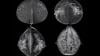
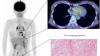
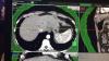
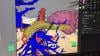
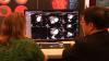
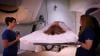
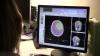
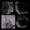
This is an example of how virtual reality is being used in neuro-radiology to better evaluate patients using advanced imaging. This dataset shows a patient's brain MRI with fused tractography imaging for pre-operative planning. This was part of a 2018 Radiological Society of North America (RSNA) hands-on session by Vinodh Kumar, M.D., and Komal Shah, M.D., associate professors of radiology at MD Anderson Cancer Center.

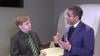
Watch the VIDEO: Using Virtual and Augmented Reality to Examine Brain Anatomy and Pathology — an interview with Vinodh Kumar, M.D., and Komal Shah, M.D.

Radiation Oncology | May 13, 2019

At ASTRO 2018, Accuray showcased new patient-first innovations, including motion synchronization on Radixact, and the new CK VoLO, a fast optimizer on the CyberKnife system. Andrew Delao, senior director of marketing for Accuray, highlights the new features.


Related content:

itnTV "Conversations": The Accuray Philosophy


Cardiac Imaging | April 26, 2019

Dee Dee Wang, M.D., director of structural heart imaging, Henry Ford Hospital, Detroit, Mich., explains how patient survival depends on keeping the left ventricular outflow track (LVOT) clear and using 3-D imaging to predict what the neo-LVOT will look like prior to transcatheter mitral valve replacement (TMVR) procedures. The close proximity between the aortic and mitral valves in the left ventricle anatomy makes it critical to assess any mitral valve overhang that will obstruct blood flow out of the left ventricle. This issue has been raised in several cardiovascular imaging structural heart intervention planning sessions at conferences over the past two years, most notably at the Society of Cardiovascular Computed Tomography (SCCT).

Read the related article Interventional Imagers: The Conductors of the Heart Team Orchestra, which Wang helped author.

Watch the related VIDEO: Overview of the Henry Ford Hospital Structural Heart Program.


Additional articles and videos on Henry Ford Hospital 

Enterprise Imaging | April 26, 2019

CarolinaEast Health System, an award-winning health system in New Bern, N.C., was one of the first to collaborate with Philips to implement IntelliSpace Enterprise Edition, a comprehensive managed service. Watch the video to see how we collaborated together to streamline workflows and improve interoperability for better care.

Watch the related editorial interview VIDEO: Streamlining PACS Administration — Interview with Mike Ciancio, imaging systems administrator at CarolinaEast Health System.


Breast Imaging | April 18, 2019

In a keynote lecture at the Society of Breast Imaging (SBI)/American College of Radiology (ACR) 2019 Symposium, Diana Miglioretti, Ph.D., dean’s professor of biostatistics at UC Davis Health, discussed risk-stratified breast cancer screening and its potential to improve the balance of screening benefits to harms by tailoring screening intensity and modality to individual risk factors.

Read the article "How Risk Stratification Might Affect Women’s Health"

Read the article "FDA Proposes New Rules for Mammography Reporting and Quality Improvement"

Watch the VIDEO: A Discussion on Proposed FDA Rules for Mammography Reporting

Information Technology | April 17, 2019

With Intellispace Enterprise Edition as the foundation, Philips Healthcare is connecting facilities and service areas within enterprises, while developing standards-based interoperability that preserves customers' investments and best of breed systems. 

Artificial Intelligence | April 17, 2019

At the Society of Breast Imaging (SBI)/American College of Radiology (ACR) 2019 Symposium, Rasu Shrestha, M.D., MBA, chief strategy officer for Atrium Health, discusses his new role with Atrium, the hype cycle of artificial intelligence (AI) and the key elements of getting AI in radiology — and in healthcare — right.

Read the article "Atrium Health Debuts Amazon Alexa Skill to Help Patients Access Medical Care"

Listen to the podcast Is Artificial Intelligence The Doom of Radiology?, a discussion with Shrestha.

Mammography | April 15, 2019

Wendie Berg, M.D., Ph.D., FACR, chief scientific advisor to DenseBreast-info.org and professor of radiology at University of Pittsburgh School of Medicine/Magee-Women’s Hospital of UPMC, spoke with ITN Editorial Director Melinda Taschetta-Millane about some of the proposed amendments to the language being used for mammography reporting and quality improvement.

Read the article "FDA Proposes New Rules for Mammography Reporting and Quality Improvement"

Information Technology | April 15, 2019

Vital Images has developed a strategy that allows its customers to capture revenues that are otherwise missed while building the infrastructure for the future. In an interview with itnTV, Vital Images executives Larry Sitka and Geoffrey Clemmons describe how the company has reconciled this vision of the future with near-term realities.

Artificial Intelligence | April 12, 2019

Paul Chang, M.D., professor of radiology, vice chair of radiology informatics and medical director for enterprise imaging, University of Chicago, explains some of the issues with artificial intelligence (AI) and how hospitals can better prepare for its eventual implementation across the field of medicine. A key takeaway is that hospitals need an infrastructure and roadway for AI and deep-learning algorithms to operate. Chang said most health systems will not invest directly in AI, but will invest in analytics, which Chang said uses much of the same infrastructure required by AI.

Chang spoke on this topic at an AIMed breakfast briefing seminar in Chicago April 9, 2019. Listen to a webcast of this hour and 15 minute talk.


Artificial Intelligence | April 02, 2019

At RSNA 2018, GE Healthcare formally presented Edison as the company's new applications platform, designed to speed the delivery of precision care. 


Related GE Edison Platform Content:

VIDEO: Artificial Intelligence - GE Builds AI Applications on Edison Platform

GE Healthcare Unveils New Applications and Smart Devices Built on Edison Platform

Advanced Visualization | April 01, 2019

GE Healthcare goes beyond core equipment maintenance to help clients solve some of their most important asset and clinical performance challenges through digital solutions.

Artificial Intelligence | March 28, 2019

GE launched a new brand that covers artificial intelligence (AI) at the Radiological Socoety of North American (RSNA) 2018 meeting. The company showed several works-in-progress, including a critical care suite of algorithms and experimental applications for brain MR. Each is being built on GE's Edison Platform.


Related GE Edison Platform Content:

GE Healthcare Unveils New Applications and Smart Devices Built on Edison Platform

VIDEO: itnTV Conversations — What is Edison?

Enterprise Imaging | March 27, 2019

GE Healthcare Centricity Clinical Archive (CCA) Analytics, shown at RSNA 2018, works directly with the vendor neutral archive (VNA), allowing users to evaluate clinical, financial and operational processes across the healthcare system. The analytics solution shows how all of the different components of the archive and all of the imaging sources — departments, facilities and modalities — are working across the enterprise.

Radiology Business | March 26, 2019

William Pinsky, M.D., a pediatric cardiologist and CEO of the Educational Commission for Foreign Medical Graduates (ECFMG), and Mandeep Mehra, M.D., medical director of the Brigham and Women’s Hospital (BWH) Heart and Vascular Center and professor of medicine at Harvard Medical School, explain the U.S. doctor shortage and how foreign doctors help fill the gap.

According to 2017 data provided by the Association of American Medical Colleges (AAMC), 40 percent of interventional cardiologists, 30 percent of cardiovascular disease specialists, and 26 percent of pediatric cardiologists in the United States are international medical graduates (IMGs). However, as the physician shortage continues to impact primary care doctors, psychiatrists, OB/GYNs, among others, the U.S. also expects to see a shortage of cardiologists within the next 10 years, according to a spotlight cardiology study issued by the professional services firm PYA, which specialized in healthcare consulting.

The interview was shot at the American College of Cardiology (ACC) 2019 meeting.


Magnetic Resonance Imaging (MRI) | March 25, 2019

Max Wintermark, M.D., chief of neuroradiology at Stanford University Medical Center, discusses the latest technologies to enhance magnetic resonance imaging (MRI) scans on display at the 2018 Radiological Society of North America (RSNA) annual meeting.

Women's Health | March 25, 2019

Deborah Levine, M.D., professor of radiology at Harvard Medical School and vice chair for academic affairs in the Department of Radiology at Beth Israel Deaconess Medical Center, Boston, describes scenarios where magnetic resonance imaging (MRI) could be more useful than ultrasound in issues with the female pelvis.

Radiology Business | March 22, 2019

American Society of Radiologic Technologists (ASRT) President Melissa Jackowski, Ed.D., R.T.(R)(M), FASRT, explains efforts by the society to have states implement licensure laws to ensure that only highly qualified RTs perform medical imaging procedures. She spoke at the Radiological Society of North America (RSNA) 2018 meeting.

Cardio-oncology | March 22, 2019

Magid Awadalla, MBBS, is an advanced cardiac imaging research fellow at Massachusetts General Hospital. He has been involved in an imaging study of cardiac changes from photon radiotherapy in breast cancer patients using serial cardiac magnetic resonance imaging (MRI). The radiotherapy beams used to treat breast cancer pass close to the neighboring heart, which can cause cardiac cell damage leading to issues like heart failure later on. He spoke on the topic of cardio-oncology at the American College of Cardiology (ACC) 2019 meeting.

Nuclear Imaging | March 22, 2019

Raza Alvi, M.D., a research fellow in radiology at Massachusetts General Hospital, has been involved in a study of a positron-emission tomography (PET) FDG radiotracer agent to image sarcoidosis. The inflammatory disease affects multiple organs and usually include abnormal masses or nodules (granulomas) consisting of inflamed tissues that can form in the heart. Alvi presented on this topic at American College of Cardiology (ACC) 2019 meeting


Related Cardiac Sarcoidosis Content:

ASNC and SNMMI Release Joint Document on Diagnosis, Treatment of Cardiac Sarcoidosis

New PET-CT Scan Improves Detection in Rare Cardiac Condition

25 Most Impactful Nuclear Cardiology Articles

Recent Advances in Cardiac Nuclear Imaging Technology

Enterprise Imaging | March 15, 2019

As a VNA, GE Healthcare Centricity Clinical Archive weaves together data from many different sources and systems. The cloud-based system, shown at RSNA 2018, offers analytics that can help physicians and administrators make decisions about the many types of data contained in this VNA, just as it can help make the VNA more efficient. 

Related content:

Artificial Intelligence | March 13, 2019

At RSNA 2018, iCad showed how its ProFound AI for digital breast tomosynthesis technology might help in the interpretation of tomosynthesis exams. Rodney Hawkins, vice president of marketing for iCad, discusses how this technology can better help detect the cancer.

Related content:

Artificial Intelligence 2018: What Radiologists Need to Know About AI

RSNA 2018 Sunday – Improving, Not Replacing

Enterprise Imaging | March 08, 2019

Mike Ciancio, imaging systems administrator at CarolinaEast Health System in North Carolina, explains how newer enterprising imaging software can improve how the backend administration of radiology PACS administration. He spoke at the 2019 Healthcare Information Management and Systems Society at (HIMSS) conference.

Ciancio said newer cloud-based systems allow for easier IT management of the backend of a PACS or enterprise imaging system.

Watch the related VIDEO: A Transformative Approach to Reducing Cost and Complexity at CarolinaEast Health System.

Look through a photo gallery of other new technologies at HIMSS19. 

Find news and videos from HIMSS 2019.

HIMSS | March 08, 2019

Karl Poterack, M.D., medical director, applied clinical informatics, Mayo Clinic, explains the role wearable devices will play in healthcare. He presented in several sessions at the 2019 Healthcare Information Management and Systems Society at (HIMSS) conference.

Poterack said there is a brewing tsunami of data in wearable technologies that healthcare systems will have to figure out how to integrate in the coming years. He said the key issue with wearable data is that there needs to be outcomes data showing the value of how many steps a patient accumulates, changes in heart rate over time, or blood pressure changes in patients with specific aliments. Without this , he said there is limited value in the information. 

Watch the related VIDEO: Use of Wearable Medical Devices for Cardiac Rehabilitation.

Look through a photo gallery of other new technologies at HIMSS19. 

Find news and videos from HIMSS 2019.

PACS | March 05, 2019

Cree Gaskin, M.D., professor, vice chair and associate chief medical officer, University of Virginia Health System, explains how new technology can be used to improve radiology reports without additional workload. His health system uses a new generation integrated RIS/PACS system that allows URL links and new graphical data presentations to be embedded into radiology reports to make them more interactive. This information can include quantifications, key images from the exam, access to full datasets, 3-D reconstructions and ability to immediately link to prior exams. He spoke is sessions on this topic at the 2019 Healthcare Information Management and Systems Society (HIMSS) conference.

Look through a photo gallery of other new technologies at HIMSS19. 

Find news and videos from HIMSS 2019.

Advanced Visualization | March 05, 2019

This is an example of a new endoscopic virtual peritoneal inflation tool on the patient's computed tomography (CT) imaging to aid in pre-procedural planning of endoscopic procedures. This is a new software feature on Fujifilm's Synapse 3D advanced visualization software released at the Healthcare Information Management and Systems Society (HIMSS) 2019 meeting. The software also enables 3-D imaging for surgical pre-procedural planning to assess the best entry points and angles. 

Look through a photo gallery of other new technologies at HIMSS19. 

Find news and videos from HIMSS 2019.

Orthopedic Imaging | March 05, 2019

This is an example of a 3-D printed pelvis that had multiple hip fractures and a second printed pelvis is from a post surgical repair CT scan, showing the pins and plates in pink. This was on display at the GE Healthcare booth at the Healthcare Information Management and Systems Society (HIMSS) 2019 conference. The files for printing were created from the CT datasets using the AW Advanced Visualization software.

Look through a photo gallery of other new technologies at HIMSS19. 

Find news and videos from HIMSS 2019.


Advanced Visualization | March 05, 2019

This is an example of a new endoscopic 3-D imaging simulator created from a patient's computed tomography (CT) scan using Fujifilm's Synapse 3D advanced visualization software. The feature was released at the Healthcare Information Management and Systems Society (HIMSS)  2019 meeting. The software enables surgical pre-procedural planning to assess the best entry points and angles. The software also enables users to perform a virtual peritoneal inflation using the advanced imaging platform.

The software shows the anatomy color coded so structures are easier to identify and to aid navigation. In this example, a pancreatic tumor is highlighted in green, which is the target of this virtual simulation. 

Find news and videos from HIMSS 2019.


Proton Therapy | March 01, 2019

ITN Associate Editor Jeff Zagoudis speaks with Mark Pankuch, Ph.D., director of physics and dosimetry at the Northwestern Medicine Chicago Proton Center, about the differences between proton and photon radiotherapy and his daily operations at the Warrenville, Ill. center.

Proton Therapy | February 28, 2019

ITN Associate Editor Jeff Zagoudis speaks with Bill Hartsell, M.D., medical director at the Northwestern Medicine Chicago Proton Center, about operating costs, reimbursement and other financial aspects of proton therapy

Watch the VIDEO: Proton Therapy Treatment at Northwestern Medicine, part 1 of the interview with Hartsell.

Additional videos and coverage of Northwestern Medicine

Information Technology | February 28, 2019

Andrew Deutsch, M.D., MBA, chairman and CEO of Renaissance Imaging Medical Associates (RIMA), an affiliate of Radiology Partners, describes RIMA’s use of an artificial intelligence (AI) based worklist workflow to manage reads across 70 sites and load balance between 120 radiologists. He spoke in sessions on this topic at the 2019 Healthcare Information Management and Systems Society at (HIMSS) conference.  

Deutsch, a nationally respected expert in skeletal radiology and magnetic resonance imaging (MRI) and in addition to his roles at RIMA, serves as medical director of the imaging departments at Northridge Hospital Medical Center and Valley Presbyterian Hospital.

Look through a photo gallery of other new technologies at HIMSS19. 

Find other news and video from HIMSS 2019. 


Related AI Coverage:

VIDEO: Editor’s Choice of the Most Innovative New Artificial Intelligence Technologies at RSNA 2018

VIDEO: Technology Report: Artificial Intelligence

How to Market Healthcare Artificial Intelligence Software

Selecting an AI Marketplace for Radiology: Key Considerations for Healthcare Providers

Cardiac Imaging | February 27, 2019

This is a virtual heart with the same electrophysiology characteristics as the real patient unveiled by Siemens at the Healthcare Information Management and Systems Society (HIMSS) 2019 annual meeting in February. This "digital twin" technology is in development and will be able to create virtual, digital organs from a patient’s medical imnaging and other physiological data. In this case, the model was created using an ECG, MRI scan and other clinical data. It was shown as a way to help optimize cardiac resynchronization therapy (CRT) lead placement. CRT currently has a 30 percent nonresponder rate, which is mainly due to the placement of leads. This model allows virtual placement of the leads In various locations to test response prior to the implantation procedure. The green dot shows the location of the virtual lead. Siemens said the technology also might have applications for testing virtual ablations strategies to save procedure time when the patient is in the EP lab

Read more about the digital twin technology.

Look through a photo gallery of other new technologies at HIMSS19. 

Find news and videos from HIMSS 2019.


Enterprise Imaging | February 27, 2019

Steve Holloway, principal analyst and company director for the healthcare market research firm Signify Research, explains the key trends he is seeing in radiology enterprise imaging systems. He spoke to ITN at the 2019 Healthcare Information Management and Systems Society at (HIMSS) conference.  

Additional HIMSS 2019 coverage.

Watch the RSNA 2018 VIDEO: Technology Report — Enterprise Imaging

Proton Therapy | February 26, 2019

ITN Associate Editor Jeff Zagoudis speaks with Bill Hartsell, M.D., medical director at the Northwestern Medicine Chicago Proton Center, about day-to-day operations and cutting-edge treatments at the Warrenville, Ill. center.

Additional videos and coverage of Northwestern Medicine

Radiation Therapy | February 21, 2019

ITN Associate Editor Jeff Zagoudis speaks with Christy Kesslering, M.D., medical director of radiation oncology at the Northwestern Medicine Cancer Center, about the different radiation therapy options for breast cancer patients offered at the center.

Watch the VIDEOs Advancements in Radiation Therapy for Brain Cancer and Multidisciplinary Treatment of Brain Tumors with Vinai Gondi, M.D., director of research and CNS neuro-oncology at the Northwestern Medicine Cancer Center.

Additional videos and coverage of Northwestern Medicine 

Radiation Therapy | February 20, 2019

ITN Associate Editor Jeff Zagoudis speaks with Vinai Gondi, M.D., director of research and CNS neuro-oncology at the Northwestern Medicine Cancer Center, about the Neuro-Oncology Multidisciplinary Clinic (NOMDC) at the center that emphasizes collaboration between clinicians to take a patient-centered approach to brain tumor cases. 

Watch the VIDEO: Advancements in Radiation Therapy for Brain Cancer, part 1 of the interview with Gondi.

Additional videos and coverage of Northwestern Medicine

Enterprise Imaging | February 20, 2019

At RSNA 2018, Philips Healthcare introduced Performance Bridge as an integral part of its IntelliSpace Enterprise Edition. The insights help drive improvements and efficiencies.

Radiation Therapy | February 15, 2019

ITN Associate Editor Jeff Zagoudis speaks with Vinai Gondi, M.D., director of research and CNS neuro-oncology at the Northwestern Medicine Cancer Center, about the cutting-edge radiotherapy treatments and research being done at the Warrenville, Ill. center.

Watch the VIDEO: Multidisciplinary Treatment of Brain Tumors, part 2 of the interview with Gondi.

Additional videos and coverage of Northwestern Medicine

Angiography | February 08, 2019

This is an example of an arterial venous malformation (AVM) in the brain imaged on a Canon Alphenix Alpha angiography system. It shjows a contrast injection highlighting the vessels, which have been color coded to show the position of the veins and arteries involved in this vascular defect. 

Read more about advances in angiography imaging systems. 

Information Technology | February 07, 2019

In this video Johann Fernando, Ph.D., Chief Operating Officer of FUJIFILM Medical Systems U.S.A., Inc. discusses his vision for Fujifilm as an end-to-end imaging partner -- offering high quality solutions for diagnostic imaging, treatment planning, treatment follow-up, and enterprise imaging management. He also shares his point-of-view on developing patient-centric solutions catering to the unique needs for specialized patient populations including solutions for pediatrics and imaging solutions dedicated to the operating room. 


Related content:

itnTV "Conversations": Collaboration as a Catalyst for AI Innovation

Fujifilm Collaborates With Lunit on AI Pilot Project

RSNA 2018 Sunday – Improving, Not Replacing

Subscribe Now