itnTV

VIDEO: One on One with Amy K. Patel, MD, American Association for Women in Radiology Immediate Past President

Breast Imaging | April 15, 2024

Don't miss ITN's latest "One on One" video interview with AAWR Past President and American College of Radiology (ACR) RAN and RADPAC Chair, Amy K. Patel, MD, discussing advocacy initiatives and innovations in artificial intelligence (AI) for breast imaging

Dr. Patel is a breast imaging trailblazer and radiology advocacy leader. In this video,  learn how radiologists can support key initiatives, ways AI is improving patient care, and more.

Related content:

Leaders from RadEqual and the AAWR Sign MOU, Solidifying Commitment to Advance Opportunities in Radiology

Technology Report: Artificial Intelligence in Radiology 2021

VIDEO: Integrating Artificial Intelligence Into Radiologists Workflow

Recent Video

Coronavirus (COVID-19) | May 07, 2020

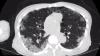
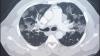
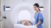
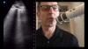
Interview with Geoffrey Rose, M.D., president of Sanger Heart and Vascular Institute with Atrium Health, in Charlotte, North Carolina, and a board member with the American Society of Echocardiography (ASE). He explains the impact if COVID-19 (SARS-CoV-2) on the cardiovascular service line and cardiac imaging. He said the virus has led to use of computed tomography (CT) not only as the frontline cardiovascular imaging modality to evaluate chest pain, but also for COVID-19 pneumonia imaging.

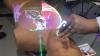
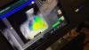
Rose said cardiac ultrasound is still used, but requires full personal protective equipment (PPE) and often abbreviated exams because of the close proximity of the sonographer and patient when performing echocardiograms. This has given rise to using dedicated point-of-care ultrasound (POCUS) systems to answer specific clinical questions quickly. Smart-phone based POCUS systems that use an app and a transducer plugged into the phone enable basic echo exams or evaluation of other parts of the anatomy quickly without the need to immediately sterilize an entire cart-based ultrasound system. These small systems also can be completely enclosed inside a transducer sheath and the phone and single transducer are much easier and faster to wipe down. He said the quality of the exams are not as good as fully enabled echocardiography systems, but it allows for quick assessments of ejection fractions and to triage if the patient needs more advanced imaging if the basic questions cannot be answered.

Since hospitals have shut down now for about two months, postponing normal checkups, and elective exams and procedures, Rose said doctors still need to visit with patients who have chronic conditions. Sanger and Atrium Heath modified its ambulatory electronic medical record (EMR) and is using video conferencing to perform virtual appointments now for the majority of these patients. He said telemedicine was not widely used before COVID-19 in his hospital system, but the pandemic will likely alter the care model for the future, with more telemedicine visits being used even after epidemic is over. He said use of POCUS and CT as frontline cardiac imaging modalities will also likely remain in place after the pandemic because of the efficiencies in care these technologies offer.


Related Coronavirus Content:

VIDEO: Imaging COVID-19 With Point-of-Care Ultrasound (POCUS)

Cardiac Imaging Best Practices During the COVID-19 Pandemic

RSNA Publishes COVID-19 Best Practices for Radiology Departments

ASE Guidelines for the Protection of Echocardiography Providers During the COVID-19 Outbreak
 

New CT Scoring Criteria for Timely Diagnosis, Treatment of Coronavirus Disease

FDA Issues New Policy for Imaging Systems During COVID-19

VIDEO: COVID-19 Precautions for Cardiac Imaging —  Interview with Stephen Bloom, M.D.

A Review of Studies Cautions Against Chest CT for Coronavirus Diagnosis


New Research Finds Chest X-ray Not Reliable Diagnostic Tool for COVID-19

VIDEO: Radiology Industry Responding to COVID-19


University of Washington Issues Radiology Policies for COVID-19

VIDEO: Best Practices for Nuclear Cardiology During the COVID-19 Pandemic — Interview with Hicham Skali, M.D.

New Research Highlights Blood Clot Dangers of COVID-19

Survey Reveals Most Medical Practices are Now Using Telehealth Due to COVID-19


CMS Offers Recommendations on Reopening Healthcare in Areas of Low COVID-19 Cases

CT Provides Best Diagnosis for Novel Coronavirus (COVID-19)

Radiology Lessons for Coronavirus From the SARS and MERS Epidemics

Radiologists Describe Coronavirus CT Imaging Features


CT Imaging of the 2019 Novel Coronavirus (2019-nCoV) Pneumonia

ACC COVID-19 recommendations for the cardiovascular care team

VIDEO: What Cardiologists Need to Know about COVID-19 — Interview with Thomas Maddox, M.D.

The Cardiac Implications of Novel Coronavirus

Coronavirus (COVID-19) | April 18, 2020

Stephen Bloom, M.D., FASNC, director of noninvasive cardiology (cardiac CT, nuclear cardiology and echocardiography) at Midwest Heart and Vascular Associates, Overland Park, Kansas. He is also a member of the American Society of Nuclear Cardiology (ASNC) Board of Directors, explains some of the issues involved and protocols used for cardiac imaging during the COVID-19 pandemic. His discussion includes computed tomography, cardiac ultrasound and nuclear imaging.

Right now, Bloom said it is difficult to test everybody and there is a shortage of masks, gowns and other personal protective equipment (PPE), and the imaging equipment needs to be sanitized each time it is used. He said it is just is not possible to image all the patients who need imaging right now. Hospitals also are trying to limit the number of healthy people people coming into hospitals for routine visits and tests to reduce their potential exposure to the novel coronavirus (COVID-19, SARS-CoV-2) and help containment efforts. 

"The tests should be done, very simply, if it changes the care of the patient. If it doesn't change the care of the patient, and it can be postponed, it should be postponed," Bloom explained. "I would say 80 percent of our cardiac imaging exams have stopped. It has been very dramatic."


Related Imaging Precautions During COVID-19 Content:

Cardiac Imaging Best Practices During the COVID-19 Pandemic

Best Practices for Nuclear Cardiology Laboratories During the Coronavirus (COVID-19) Pandemic

ASE Guidelines for the Protection of Echocardiography Providers During the COVID-19 Outbreak 

VIDEO: Best Practices for Nuclear Cardiology During the COVID-19 Pandemic — Interview with Hicham Skali, M.D.

VIDEO: Cancelling Non-essential Cardiac Procedures During the COVID-19 Outbreak — Interview with Ehtisham Mahmud, M.D. 

VIDEO: 9 Cardiologists Share COVID-19 Takeaways From Across the U.S.  

VIDEO: Telemedicine in Cardiology and Medical Imaging During COVID-19 — Interview with Regina Druz, M.D.

VIDEO: Use of Teleradiology During the COVID-19 Pandemic — an interview with radiologist John Kim, M.D.

Study Looks at CT Findings of COVID-19 Through Recovery

Experts Stress Radiology Preparedness for COVID-19

VIDEO: Imaging COVID-19 With Point-of-Care Ultrasound (POCUS) — Interview with emergency physician Mike Stone, M.D.,

VIDEO: How China Leveraged Health IT to Combat COVID-19 — Interview with Jilan Liu, M.D., CEO for the HIMSS Greater China

ACR Recommendations for the Use of Chest Radiography and CT for Suspected COVID-19 Cases

VIDEO: What Cardiologists Need to Know about COVID-19 — Interview with Thomas Maddox, M.D.

The Cardiac Implications of Novel Coronavirus

Computed Tomography (CT) | April 09, 2020

Jeffrey Bundy, Ph.D., chief executive officer, United Imaging in the United States, explains the company's efforts to respond to the novel coronavirus (COVID-19) pandemic in the U.S., Europe and China. The company is putting its first semi-trailer based computed tomography (CT) system into service at a hospital in New York City.

United Imaging began working on new solutions for the U.S. after getting requests from its customers and seeing the needs in China and Europe. The company is collaborating with different vendors, including Sea Box and AMST to build both semi-truck trailer CT scanning rooms and those built into customized shipping containers. Other vendors are helping with infection control, ventilation, and easier cleaning/disinfection to help protect healthcare workers and patients.

The first transportable CT scanner was installed at the request of Maimonides Medical Center, the largest hospital in Brooklyn, New York, on the front lines of the current crisis. As the hospital scales its operations to meet the needs of an expected influx of coronavirus patients, doubling its capacity to 1,400 beds, United Imaging’s scanner will help expand its capacity for imaging studies to support diagnosis and treatment.

United Imaging Confirms Rapid Deployment of Transportable CT Scanners

United Imaging Sends Out More than 100 CT Scanners and X-ray Machines to Aid Diagnosis of the Coronavirus


Coronavirus (COVID-19) | April 04, 2020

Hicham Skali, M.D., a staff cardiologist and member of the Non-invasive Cardiovascular Imaging Program at Brigham and Women’s Hospital (BWH), and at Brigham and Women’s / Massachusetts General Health Care Center at Foxborough, explains the new recommendations from the American Society of Nuclear Cardiology (ASNC) and from imagers in China and Singapore. The ASNC created a best practices document for nuclear cardiology laboratories during the novel coronavirus (COVID-19, SARS-CoV-2) pandemic. The suggestions in the guidelines can ally to any imaging modality, including computed tomography (CT), MRI and ultrasound. 

Skali elaborates on the following points in his discussion, which are specific recommendations in the ASNC and SNMMI COVID-19 guidance document:
   • Rescheduling non-urgent visits
   • Rescheduling elective surgeries and procedures
   • Using separate spaces for patients with known or suspected COVID-19 to prevent spread
   • Ensuring supplies are available
   • Promoting use of telehealth
   • Screen staff, patients and visitors before they enter the department
   • Minimize non-essential visitors into the department
   • Record symptoms at the start of the shift
   • Use personal protective equipment (PPE)for healthcare personnel
   • If available, use PPE for patients due to concern of asymptomatic transmission of COVID-19
   • Maintain strict hand hygiene
   • Maintain 6 feet distance in all patient/staff interactions when possible
   • Work remotely whenever feasible, especially with ready studies
   • Rotating staff schedules for on-site and off-site work
   • Use of rest only studies if possible
   • Use of half-time SPECT to speed exam times
   • Use of PET if available to speed exam times

Skali served as the moderator in for the ASNC on demand webinar COVID-19 Preparedness for Nuclear Cardiology Labs: Insights from the US, China and Singapore.

VIDEO: Telemedicine in Cardiology and Medical Imaging During COVID-19 — Interview with Regina Druz, M.D., an ASNC Board member and also a speaker during the ASNC webinar.

Find more news and video on relating to COVID-19 and its impact on radiology

Coronavirus (COVID-19) | March 27, 2020

Regina Druz, M.D., FASNC, a member of the American Society of Nuclear Cardiology (ASNC) Board of Directors, chairwomen of the American College of Cardiology (ACC) Healthcare Innovation Section, and a cardiologist at Integrative Cardiology Center of Long Island, N.Y., explains the rapid expansion of telemedicine with the U.S. spread of novel coronavirus (COVID-19, SARS-CoV-2).

Druz spoke on the unprecedented expansion of telemedicine in the U.S. under COVID-19, seeing more use in the last two months, as opposed to the past two decades. The Centers for Medicare and Medicaid Services (CMS) previously only reimbursed for Telehealth in rural areas it determined had a shortage of doctors. However, in early March 2020, CMS dropped the geographic requirements and allowed Telehealth usage across th country as a way to mitigate person-to-person contact and keep vulnerable, older patients at home for routine check ups with doctors.

Druz has subspecialty certifications in nuclear cardiology, adult echocardiography and cardiac computed tomography (CT) and explains how Telehealth can be used to pre-screen patients and get patient sign off on procedures prior to coming in for an exam, helping speed the process in the hospital and limit personal contact.

Concerns about the rpaid spread of COVID-19 also has driven many radiology departments to convert to wider use of teleradiology to allow more radiologists to work from home and reduce person-to-person contact within the hospitals. 

Watch the related VIDEO: Use of Teleradiology During the COVID-19 Pandemic — an interview with John Kim, M.D., chairman, Department of Radiology, THR Presbyterian Plano, Texas, and chief technology officer at Texas Radiology Associates.


Related COVID-19 Content:

VIDEO: Imaging COVID-19 With Point-of-Care Ultrasound (POCUS) — Interview with emergency physician Mike Stone, M.D.,

VIDEO: How China Leveraged Health IT to Combat COVID-19 — Interview with Jilan Liu, M.D., CEO for the HIMSS Greater China

Study Looks at CT Findings of COVID-19 Through Recovery

Experts Stress Radiology Preparedness for COVID-19

ACR Recommendations for the Use of Chest Radiography and CT for Suspected COVID-19 Cases

VIDEO: What Cardiologists Need to Know about COVID-19 — Interview with Thomas Maddox, M.D.

The Cardiac Implications of Novel Coronavirus


Teleradiology | March 26, 2020

John Kim, M.D., chairman, Department of Radiology, THR Presbyterian Plano, Texas, and chief technology officer at Texas Radiology Associates, explains the use of teleradiology during the novel coronavirus (COVID-19) outbreak in the U.S. The radiology group is part of Collaborative Imaging, a radiologist owned teleradiology alliance that enables radiologists to read studies using secure web links and an app.

The rapid spread of coronavirus has increased the value telemedicine solutions to help prevent person-to-person contact and take some clinicians out of the hospitals to work at home or office locations. This includes the use of teleradiology tools to read images and communicate with referring physicians.

Telemedicine allows for the continuity of care within healthcare facilities and patients and allows for safe distance without sacrificing the quality of care. It also saves patients from spreading germs to others, like while on public transportation or in the doctor’s waiting room, and to the healthcare providers who tend to them. Televisits also only last an average of 10 minutes, allowing physicians to assess patients more quickly and easily. 

Coronavirus (COVID-19) | March 25, 2020

Jilan Liu, M.D., MHA, CEO for the HIMSS Greater China, explains how health information technology (HIT) was leveraged in China to help fight the outbreak of novel coronavirus (COVID-19, SARS-CoV-2). Her discussion includes use of artificial intelligence, telemedicine to reduce contact between people, using the electronic medical record to help record and track people COVID-19 patients had contact with, apps, internet hospitals, and how 5G networks helped speed up IT systems.

She was previously principal and consulting director of the Greater China Joint Commission International (JCI). As chief executive for HIMSS Greater China, Liu and her team made a significant breakthroughs with hospitals and the HIT industry in the greater China region to embrace international HIT standards, certification requirements and to rethink prudent approaches to IT investments and operations. 

Read the the related article by Liu, "Deployment of Health IT in China’s Fight Against the COVID-19 Epidemic."


Ultrasound Imaging | March 20, 2020

Interview with Mike Stone, M.D., an emergency physician at Northwest Acute Care Specialists in Portland, Ore., director of point of care ultrasound education at Butterfly Network Inc., and and former chief of the division of emergency ultrasound at Brigham and Women's Hospital, Boston. He explains what clinicians look for in lung ultrasound to evaluate patients for plural wall thickening and areas of congestion inside the lung typical of pneumonia and seen in patients with novel coronavirus (COVID-19, also referred to clinically as SARS-COV-2). 

Small hand-held ultrasound systems may offer advantages over X-ray or computed tomography (CT), because the entire system can be contained inside a protective sheath, making it easier to disinfect the device rather than an entire imaging room. Point of care ultrasound (POCUS) systems also allow triage of patients in tents or other areas outside of the hospital where other imaging modalities are not available. 

Stone shares examples of COVID-19 ultrasound images from Butterfly Network POCUS system technology, which turns an iPhone into an FDA-cleared ultrasound device for multiple POCUS exam types. It is the first POCUS system reported to be used for triage and monitoring COVID-19 patients. 
 

Related Coronavirus Content:

Study Looks at CT Findings of COVID-19 Through Recovery

Experts Stress Radiology Preparedness for COVID-19

ACR Recommendations for the Use of Chest Radiography and CT for Suspected COVID-19 Cases

Wuhan CT Scans Reliable for Coronavirus Diagnosis, Limited for Differentiation

The Cardiac Implications of Novel Coronavirus

CT Provides Best Diagnosis for Novel Coronavirus (COVID-19)

Radiology Lessons for Coronavirus From the SARS and MERS Epidemics

Deployment of Health IT in China’s Fight Against the COVID-19 Epidemic

Emerging Technologies Proving Value in Chinese Coronavirus Fight

Radiologists Describe Coronavirus CT Imaging Features

Coronavirus Update from the FDA

CT Imaging of the 2019 Novel Coronavirus (2019-nCoV) Pneumonia

CT Imaging Features of 2019 Novel Coronavirus (2019-nCoV)

Chest CT Findings of Patients Infected With Novel Coronavirus 2019-nCoV Pneumonia 

VIDEO: What Cardiologists Need to Know about COVID-19 — Interview with Thomas Maddox, M.D.

The Cardiac Implications of Novel Coronavirus

ESC Council on Hypertension Says ACE-I and ARBs Do Not Increase COVID-19 Mortality

Find more related clinical content Coronavirus (COVID-19)


Additional COVID-19 Resources for Clinicians:

   ACC COVID-19 Hub page   

   Johns Hopkins Coronavirus Resource Center with inteavtive map of cases in U.S. and worldwide 

   World Health Organization (WHO) COVID-19 situation reports

   World Health Organization (WHO) coronavirus information page

   U.S. Food and Drug Administration (FDA) COVID-19 information page

   Centers for Disease Control (CDC) COVID-19 information page

   Centers for Medicare and Medicaid Services (CMS) frequently asked questions and answers (FAQs) for healthcare providers regarding COVID-19 related payments

Enterprise Imaging | March 02, 2020

At RSNA19, Philips discussed with ITN Contributing Editor Greg Freiherr how its IntelliSpace Enterprise Edition fits into the Philips portfolio, and how the corporate acquisition of Carestream Health IT might affect the company. The video also discusses how Philips will help prospective customers acquire its products.

Artificial Intelligence | February 21, 2020

In Artificial Intelligence at RSNA 2019, ITN Contributing Editor Greg Freiherr offers an overview of artificial intelligence (AI) advances at the Radiological Society of North America (RSNA) 2019 annual meeting.

Enterprise Imaging | February 21, 2020

In Enterprise Imaging at RSNA 2019, ITN Contributing Editor Greg Freiherr offers an overview of enterprise imaging advances at the Radiological Society of North America (RSNA) 2019 annual meeting.

Enterprise Imaging | February 19, 2020

Bill Lacy, vice president, Medical Informatics at FUJIFILM Medical Systems U.S.A., Inc. discusses Synapse 7x, a convergence of the company’s server-side technology designed to cover all the different areas of diagnostic visualization, as well as overall enterprise viewing, with ITN Consulting Editor Greg Freiherr. For more information on Fujifilm's Enterprise Imaging solutions, visit ei.fujimed.com

Flat Panel Displays | February 19, 2020

EIZO medical monitors were showcased recently at RSNA 2019. Learn how the EIZO approach streamlines the effectiveness of your readings. From the NEW 30.9 inch, super-high-resolution, 12 megapixel multi-modality Radiforce RX1270, to the EIZO built-in features such as Point-and-Focus, EIZO holds to the ideal to harmoniously combine performance, ease-of-use and comfort. Learn the Shape of Comfort with EIZO.  Stop by and visit with us at HIMSS, booth #4087.

Artificial Intelligence | February 07, 2020

At RSNA19, GE Healthcare introduced its Edison Open AI Orchestrator. The software has been designed to operate smart algorithms that might save radiologists time. ITN Contributing Editor Greg Freiherr discusses its benefits with Karley Yoder, vice president and general manager of artificial intelligence for GE.


Related GE Edison Platform Content:

GE Healthcare Unveils New Applications and Smart Devices Built on Edison Platform

VIDEO: itnTV Conversations — What is Edison?

Artificial Intelligence | February 06, 2020

ProFound AI is an FDA-cleared artificial intelligence (AI) system for reading 3-D breast tomosynthesis images. At RSNA19, ITN Contributing Editor Greg Freiherr spoke with iCad Chairman and CEO Michael Klein about the system, which has been clinically proven in a large reader study to produce an 8% average improvement in sensitivity, 7.2% average reduction in recall rate and 52.7% reduction in average radiologist reading time. 

Mammography | January 24, 2020

Imaging Technology News Contributing Editor Greg Freiherr interviewed Henry Izawa, vice president of modality solutions, Fujifilm Medical Systems U.S.A., Inc. about Fujifilm's latest innovations in mammography.

In this video, Izawa announces the latest imaging advancements available for the Aspire Cristalle digital mammography system with digital breast tomosynthesis, including S-View, a synthesized 2-D image reconstructed from DBT and other patient enhancements such as the comfort paddle to provide a noticeably more comfortable exam for the patient.

Watch the video to hear all about Fujifilm's updates.

To learn more, visit www.fujimed.com

Enterprise Imaging | January 20, 2020

GE Healthcare's iCenter is a cloud-based management software that provides 24/7 visibility to customers' visual and operational data. In this Conversations video, Contributing Editor Greg Freiherr discusses iCenter with GE and Microsoft Executives at RSNA 2019.

RSNA | January 13, 2020

ITN Editor Dave Fornell takes a tour of some of the most innovative new medical imaging technologies displayed on the expo floor at the Radiological Society of North America (RSNA) 2019 meeting. 

Technology examples include a robotic arm to perform remote ultrasound exams, integration of artificial intelligence (AI) to speed or automate radiology workflow, holographic medical imaging display screens, a new glassless digital radiography (DR) X-ray detector, augmented reality for transesophageal echo (TEE) training, moving DR X-ray images, 3-D printed surgical implants created from a patient's CT imaging, DR X-ray tomosynthesis datasets, radiation dose management and analytics software, and new computed tomography (CT) technologies.

Photo Gallery of New Imaging Technologies at RSNA 2019

Find more videos and news from RSNA 2019


RSNA | January 07, 2020

Elizabeth M. Hecht, M.D., professor of radiology, Columbia University, New York, explains the latest advances to help visualize the difficult to image pancreas. She was a moderator of a session on pancreatic imaging advances at the 2019 Radiological Society of North America (RSNA) meeting.

She explained computed tomography is the front line imaging modality, followed by MRI for more detailed examination of the soft tissue details. MRI tissue elastography can also help to differentiate pancreatitis from pancreatic cancer. Hecht also said PET-CT and endoscopic ultrasound also play roles in pancreatic imaging. She said new software is helping automate measurements and artificial intelligence (AI) is in development to help improve pancreatic imaging and to find new radiomic markers that might be missed by the human eye to better detect disease or risk stratify patients.

Find more news and video from RSNA


Magnetic Resonance Imaging (MRI) | January 06, 2020

Karen Ordovas, M.D., MAS, professor of radiology and cardiology at the University of California San Francisco (UCFS) School of Medicine and a Society of Cardiac Magnetic Resonance (SCMR) board member, explains how cardiac MRI can help in women's heart disease and to better define complex congenital heart anatomy. She spoke at the 2019 Radiological Society of North America (RSNA) meeting. 

She specializes in cardiac and pulmonary imaging, and has particular expertise in using CT and MRI techniques in cardiovascular imaging and the differences of presentation in imaging between male and female cardiac patients. Ordovas is helping advance education around heart disease in women and bring great awareness of quality tools to diagnose heart disease and how heart MRI can help. She also is heavily involved in the use of heart MRIs for pregnant women, since there is no radiation,  and patients with congenital heart disease where detailed imaging of the complex anatomy is required.

The use of cardiac MRI in congenital heart disease is common in serial imaging of patients with Tetralogy of Fallot (TOF), one of the most common congenital heart diseases for which patients are referred for post-operative magnetic resonance (MR) imaging evaluation. In the past few decades, surgery has proved successful, but most patients require repeat imaging throughout their lives and MRI can offer more detailed soft tissue imaging without the use of radiation. 


Related Cardiac MRI Content:

VIDEO: Advances in Cardiac MRI Technology — Interview with James Carr, M.D.

Cardiac MRI Delivers Accurate Diagnosis for Frontline Chest Pain Evaluation

VIDEO: Dedicated Cardiac MRI Use at the Baylor Scott White Heart Hospital

Advantages and New Applications of Cardiac MRI

Will Cardiac MRI Expand?


Digital Radiography (DR) | January 06, 2020

An experienced technologist and two Agfa executives talk about what distinguishes the new Agfa 100s. Three key highlights include the quality of images that users can get out of the system for the certainty of the diagnostic; more flexibility that has been designed around the user; and its revolutionary platform.

Ultrasound Imaging | January 06, 2020

The Arietta 850SE provides facilities with numerous features and functionality to get the most out of a system. The CMUT probe performs the role of multiple probes while the single-crystal C252 probe gets increased penetration. eFocusing simplifies scanning techniques and shearwave elastography provides unique feedback on tissue stiffness.

Computed Tomography (CT) | January 06, 2020

Hitachi announced the FDA clearance of its newest CT – Scenaria View – at RSNA2019. The View offers an 80 cm wide bore, 550 lb table capacity and powerful 84 kW generator as well as a 200 mm lateral shifting table. Installs in Georgia, Ohio and Florida are underway with more to come in the new year.

Digital Radiography (DR) | January 03, 2020

At RSNA19, David Widmann, president and CEO of Konica Minolta Healthcare Americas, discussed innovation, stressing the importance of academic relationships. 

Computed Tomography (CT) | January 03, 2020

In this Conversations video at AHRA, Ryan K. Lee, M.D., describes the harm extravasation can do and why using power injectors to administer saline beforehand increases patient safety based on his experiences at the Einstein Healthcare Network. 

Stroke | January 03, 2020

Ajay Choudhri, M.D., chairman of radiology, Capital Health, Hopewell, N.J., explains his center's experience using an artificial intelligence (AI) application to help auto detect intracranial hemorrhage. There are several AI stroke auto detection apps now available with FDA clearance or in development that were shown at the Radiological Society Of North America (RSNA) 2019 meeting. These are being adopted by hospitals and multi-center radiology practices the U.S. to flag suspected cases of ischemic stroke or brain bleeds for immediate reads. 

MaxQ AI Accipio hemorrhagic stroke detection software. MaxQ.AI
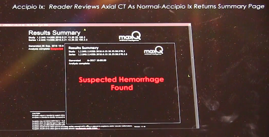
The software also offers a second set of eyes for more difficult to detect cases. Quickly determining is a stroke is ischemic or hemorrhagic is critical to the path of treatment. If caught early enough, TPA can be injected into patients to clear clots causing an ischemic stroke, but can cause massive brain damage or death if injected into a patient with a brain bleed. At advanced neuro-interventional centers, quickly determining the type of stroke is needed to know if they need to revascularize a patient or manage a hemorrhage. 


Related Content:

How Artificial Intelligence Can Predict and Detect Stroke

MaxQ AI's Intracranial Hemorrhage Software to be Integrated on Philips CT Systems

Find more news and video from RSNA

Orthopedic Imaging | January 03, 2020

This is a demo of the EOS orthopedic X-ray imaging system at the recent 2019 Radiological Society of North America (RSNA) meeting. The system allows for weight bearing exams. EOS Imaging System the first technology capable of providing full-body, 2-D and 3-D images of pediatric patients in a standing position at a low dose of radiation. The vendor said the scans are much lower dose than computed tomography (CT).

EOS captures bi-planar images with two perpendicular X-ray beams that travel vertically while scanning the patient from head to toe. In less than 20 seconds, the EOS exam produces simultaneous frontal and lateral, low dose images. The two resulting digital images are processed by EOS' proprietary sterEOS software to generate a 3-D model of the patient's spine and/or lower limbs. These detailed images with only 20 seconds of radiation were previously unachievable before EOS technology.

Read more about the system and a recent install

VIDEO: Editors Choice of the Most Innovative New Radiology Technology at RSNA 2019

Photo Gallery of New Imaging Technologies at RSNA 2019

Find more news and video from RSNA 2019


Advanced Visualization | December 30, 2019

This is a hologram of a fracture from a computed tomography (CT) scan displayed by the start up company Voxon at the 2019 Radiological Society Of North America (RSNA) meeting. The technology uses a half millimeter thick glass plate that pulses up and down very rapidly while projecting 4,000 images per second. It can display standard DICOM radiology files or STL files used for 3-D printing.

There were at least four vendors showing holographic screens to display advanced visualization 3-D renderings of anatomy from medical imaging. All four of these screens could be viewed in true 3-D using normal vision without the need for special glasses or a virtual reality visor.

The images in this example flickers because of the different frame rates of the system and the iPhone used to film it, but the actual images appears much more stable.

This technology was also included in the VIDEO: Editors Choice of the Most Innovative New Radiology Technology at RSNA 2019

Photo Gallery of New Imaging Technologies at RSNA 2019

Find more news and video from RSNA


MRI Breast | December 26, 2019

Gillian Newstead, M.D., director of global breast imaging and former professor at the University of Chicago, explains a large breast MRI screening study presented at the 2019 Radiological Society of North America (RSNA) meeting. 

The DENSE Trial Study Group included 40,000 women from the Netherlands who underwent standard mammography. Of these, 8,061 women were identified as having dense breasts and were offered supplemental MRI screening, and about half took advantage of the additional screening. Dense breast tissue which can obscure cancer on conventional mammograms but not on MRI. The MRI cancer-detection rate among the women who actually underwent MRI screening was 16.5 per 1,000 screenings.

The use of supplemental magnetic resonance imaging (MRI) screening in women with extremely dense breast tissue and normal results on mammography resulted in the diagnosis of significantly fewer interval cancers than mammography alone during a two-year screening period. The results were published in the Nov.  28, 2019, issue of The New England Journal of Medicine and presented earlier at the European Congress of Radiology (ECR) 2019.


Related Breast MRI and Dense Breast Content:

Breast Density Explained

Animation to Bring Clarity to Dense Breasts

Improving Clinical Image Quality for Breast Imaging

Breast Imaging in the Age of Coronavirus

Abbreviated MRI Outperforms 3-D Mammograms at Finding Cancer in Dense Breasts

VIDEO: Explaining Dense Breasts — Interview with Christiane Kuhl, M.D.

Technologies to Watch in Breast Imaging

Screening MRI Detects BI-RADS 3 Breast Cancer in High-risk Patients
 

Reference:

Bakker MF, de Lange SV, Pijnappel RM, et al. Supplemental MRI Screening for Women with Extremely Dense Breast Tissue. N Engl J Med 2019; 381:2091-2102. DOI: 10.1056/NEJMoa1903986.

Computed Tomography (CT) | December 26, 2019

Kevin Little, Ph.D., assistant professor and medical physicist, Department of Radiology, at The Ohio State University, explains how hospitals can better manage their computed tomography (CT) imaging protocols across their fleet of CT scanners from various vendors. He moderated a session on this topic at the 2019 Radiological Society of North America (RSNA) meeting.

He stressed the importance of CT protocol management to an imaging practice and showed examples of tools that can be used to develop consistent protocols across multiple systems. Little said it is important to develop awareness of the Management of Acquisition Profiles (MAP) IHE profile and features that should be requested from CT scanner vendors.

CT protocols, which include all clinical and technical parameters for a given study, are the starting point for achieving high-quality images with reasonable radiation and contrast doses. An imaging practice that desires to follow the "as low as reasonably achievable” (ALARA) principle and produce high-quality images should have standardized protocols across their enterprise. Accreditation standards require a periodic review of all scanner protocols. However, variations among vendors, models, and clinical indications mean that managing and optimizing dozens of parameters for each protocol on every scanner in a health system is challenging. Even when variations between systems are limited, managing protocol names and parameters across multiple systems can be difficult. The purpose of this symposium is to identify tools and techniques that may be used to manage protocols across multiple systems and to provide a framework for protocol optimization.

Magnetic Resonance Imaging (MRI) | December 20, 2019

James Carr, M.D., chair of the Department of Radiology, Northwestern University, and incoming 2020 President of the Society of Cardiac Magnetic Resonance (SCMR), explains why MRI is an ideal cardiac imaging modality, at the 2019 Radiological Society of North America (RSNA) meeting.

Heart MRI offers advantages over computed tomography (CT) and echocardiography because of its excellent soft tissue delineation and its ability to offer information beyond anatomical imaging, such as perfusion, morphology and metabolism. MRI can be technically challenging and the exams requiring a long time, but recent advances have helped cur cardiac imaging times down significantly. Automation and artificial intelligence (AI) also is making post-processing and quantification mush faster, brining it closer to the time it takes to scan and post-process CT imaging.
 
Northwestern is was one of the early adopters of cardiovascular MRI. Carr said heart MRI was not common in regular clinical use until the past decade at some luminary centers. In 2005, Carr was given the opportunity to develop a clinical cardiac MRI program at Northwestern.
 
He said MRI scanners have improved, and now much faster than a decade ago. They are also more optimized for cardiac imaging. While heart MRI is well known in large hospital centers, Carr said it still needs to develop and expand to community hospitals and rural hospitals outside major population centers. 

Artificial intelligence is playing a significant role in cardiac MRI automation of speeding workflow and quantification. Carr said these technologies will become mainstream in the next few years. AI also will play an increasing role in risk prediction based on new image analysis algorithms in development.

For more information on cardiac MRI, visit SCMR's website www.heartmri.org.

Radiation Dose Management | December 19, 2019

Mahadevappa Mahesh, Ph.D., chief of medical physicist and professor of radiology and medical physics, Johns Hopkins University, Baltimore, treasurer of the American Association of Physicists in Medicine (AAPM),a board member of the American College of Radiology (ACR), presented a late-breaking study on how medical imaging radiation dose has started to drop over the past decade. He is the co-chair of the National Council on Radiation Protection and Measures Report (NCRP), and presented the most recent NCRP data analysis at the 2019 Radiological Society of North America (RSNA) meeting.

The new NCRP 184 report covers the period between 2006 and 2016, the period of the most current CMS data. It shows a decrease of about 20 percent in the radiation dose the U.S. population receives from medical imaging, compared to the NCRP 160 that covered the period of up to 2006.

Key findings of the study include:

   • CT dose dropped about 6 percent, despite a 20 percent increase CT scans since 2006;

   • Drop of more than 50 percent for nuclear imaging scans, mainly due to fewer procedures being performed;

   • A 15-20 percent decrease across X-ray imaging modalities.

Mahesh says this shows the impact of using "as low as reasonably achievable" (ALARA) principals, new dose guidelines outlined jointly by numerous medical societies, and dose reduction initiatives like Image Wisely, Image Gently, and the American College of Radiology (ACR) Dose Index Registry.

He said there was growing concern a decade ago when the last council report was published, which showed a steep increase in radiation dose. This was mainly due to a rapid increase in the use of computed tomography (CT) and other types of X-ray based and nuclear radiotracer medical imaging. This prompted the ACR to create the Image Wisely program and push for the use of more thoughtful imaging doses based on patient size, using the "as low as reasonably achievable” (ALARA) principle. While CT dose was lowered, he said the biggest decline over all was in nuclear imaging.


Related Medical Imaging Radiation Dose Resources:

VIDEO: Radiation Dose Monitoring in Medical Imaging — an interview with Mahadevappa Mahesh, Ph.D.

The Basics of Radiation Dose Monitoring in Medical Imaging

How to Understand and Communicate Radiation Risk — Image Wisely

Radiation in Medicine: Medical Imaging Procedures

FDA White Paper: Initiative to Reduce Unnecessary Radiation Exposure from Medical Imaging

Radiation Dose in X-Ray and CT Exams

Radiation Dose from Medical Imaging: A Primer for Emergency Physicians

Radiation risk from medical imaging

FDA: Medical X-ray Imaging


Find RSNA news and other videos


Ultrasound Transesophageal echo (TEE) | December 19, 2019

This is an example of an augmented reality (AR) training system for transesophageal echo (TEE) created by the simulation company CAE. Rather than just looking at an overhead screen, this system allows the user to use a HoloLens visor to see the impact their probe manipulation has on the cardiac ultrasound imaging and better shows the orientation of the ultrasound probe, the 2-D ultrasound image slice and the relation to the anatomy. It was displayed at the 2019 Radiological Society Of North America (RSNA) meeting.

Read more about this technology.

Find more technology news and video from the RSNA meeting

RSNA | December 18, 2019

ITN Editor Dave Fornell and ITN Consulting Editor Greg Freiherr offer a post-game report on the trends and technologies they saw on the expo floor of 2019 Radiological Society of North America (RSNA) annual meeting. This includes artificial intelligence (AI), augmented reality, holographic imaging, cybersecurity and advances in digital radiography (DR) with a glassless detector plate, X-ray tomosynthesis, dual-energy X-ray and dynamic DR imaging. 

VIDEO: Editors Choice of the Most Innovative New Radiology Technology at RSNA 2019

Photo Gallery of New Imaging Technologies at RSNA 2019

Find RSNA news and other videos


Proton Therapy | December 16, 2019

Join Chris Toth, president of Varian’s Oncology Systems business, for a peek at the history of machine learning/AI in radiation oncology, plus other highlights in 2019:

  • Ethos therapy: the world’s first AI-powered adaptive radiotherapy.
  • Noona cloud-based application for capturing patient-reported outcomes.
  • Varian’s multi-room configuration for ProBeam 360 proton therapy.
  • The promise of FLASH, an ultra-high-speed treatment that is in pre-clinical testing, and represents an exciting and potentially promising new direction in the treatment of cancer. 
Digital Radiography (DR) | December 12, 2019

Vieworks to present NEW DR detectors, VIVIX-S V series. This is a strategic model that will open up a new era of digital radiography. VIEW it now. You will know.

Quality Assurance (QA) | December 06, 2019

Modus QA is proud to offer a superior phantom for quantifying geometric distortion in modern MRgRT applications. Watch this video to discover how the entire QA process including set-up and data analysis can be completed in under 10 minutes.

Artificial Intelligence | October 22, 2019

David Sjostrom, Ph.D., deputy chief physicist, Herlev Hospital, Department of Oncology, Division of Radiotherapy, Herlev, Denmark, shares the first clinical experience treating cancer patients with the Varian Ethos radiation therapy system. He spoke to ITN at ASTRO 2019, where he presented information on the first 5 patients in the world being treated with this new technology. It uses artificial intelligence to take the onboard cone beam CT scans to automatically create an adaptive plan for any changes in patient weight loss, bladder volume, or change in tumor size. The plan can be available in minutes while the patient is on the table. It enables sparing of more healthy tissue and makes adaptive therapy much easier to use. 


Radiation Oncology | October 11, 2019

Lorraine Drapek, DNP, nurse practitioner, radiation oncology, GI service, Massachusetts General Hospital, explains the roles of advanced practice providers in radiation therapy. She spoke on this topic at ASTRO 2019 at a session that reviewed the integration of APPs into radiation oncology practice to enhance clinical care. This includes but is not limited to: on-treatment management, symptom and acute toxicity management during treatment, inpatient consultations, procedural assistance, treatment planning, follow-up, survivorship and research.

Radiation Therapy | October 08, 2019

Kristin Higgins, M.D., medical director of radiation oncology, Emory Clinic at the Winship Cancer Institute, explains considerations when treating previous radiation oncology patients again at the same or other tumor sites. She spoke on this topic at the American Society for Radiation Oncology (ASTRO) 2019 annual meeting in Chicago.

More coverage of ASTRO 2019

Subscribe Now