itnTV

VIDEO: One on One with Amy K. Patel, MD, American Association for Women in Radiology Immediate Past President

Breast Imaging | April 15, 2024

Don't miss ITN's latest "One on One" video interview with AAWR Past President and American College of Radiology (ACR) RAN and RADPAC Chair, Amy K. Patel, MD, discussing advocacy initiatives and innovations in artificial intelligence (AI) for breast imaging

Dr. Patel is a breast imaging trailblazer and radiology advocacy leader. In this video,  learn how radiologists can support key initiatives, ways AI is improving patient care, and more.

Related content:

Leaders from RadEqual and the AAWR Sign MOU, Solidifying Commitment to Advance Opportunities in Radiology

Technology Report: Artificial Intelligence in Radiology 2021

VIDEO: Integrating Artificial Intelligence Into Radiologists Workflow

Information Technology

Artificial Intelligence | December 02, 2020

Kirti Magudia, M.D., Ph.D., an abdominal imaging and ultrasound fellow at the University of California San Francisco, explains how an automated deep learning analysis of abdominal computed tomography (CT) images can produce a more precise measurement of body composition and better predicts major cardiovascular events, such as heart attack and stroke, better than overall weight or body mass index (BMI). This was according to a study she presented at the 2020 Radiological Society of North America (RSNA) virtual meeting.

Unlike BMI, which is based on height and weight, a single axial CT slice of the abdomen visualizes the volume of subcutaneous fat area, visceral fat area and skeletal muscle area. However, manually measuring these individual areas is time intensive and costly. A multidisciplinary team of researchers, including radiologists, a data scientist and biostatistician, developed a fully automated artificial intelligence (AI) method to determine body composition metrics from abdominal CT images.

Statistical analysis demonstrated that visceral fat area was independently associated with future heart attack and stroke. BMI was not associated with heart attack or stroke. 

Read more about this study

Find more RSNA news

Information Technology | December 01, 2020

Treating cancer effectively often includes a combination of patient therapies. In recent years, technology advancements have led to a more efficient and personalized approach to treatment. Andrew Wilson, President of Oncology Informatics at Elekta, discussed the latest software advancements with ITN.

Remote Viewing Systems | November 28, 2020

Konica Minolta’s theme for RSNA 2020 is Depth of Vision. ITN recently talked with David Widmann, President and CEO of Konica Minolta Healthcare Americas, about this focus and their key messages for customers and RSNA attendees.

X-Ray | November 28, 2020

Agfa is looking to transform X-ray with new advancements in volumetric imaging, and with new mobile concepts and implementation of intelligent tools. ITN had a conversation with Georges Espada on Transforming X-ray with Intelligent Tools. 

Enterprise Imaging | November 23, 2020

Fujifilm's next generation secure server-side viewer platform extends across enterprise imaging areas to bring together radiology, mammography and cardiology into a single zero footprint platform. Bill Lacy, vice president of medical informatics for Fujifilm Medical Systems USA recently talked with ITN about their Synapse 7x platform.

Artificial Intelligence | November 11, 2020

Artificial Intelligence (AI) is becoming more common place in radiology practices, and emerging technologies are providing radiologists with sophisticated detection software to aid their reading and provide support for a busy workflow. With the progression of AI technology, vendors must look not only at what AI can do for the radiologist, but how the radiologist and the technician interact with that technology –  the goal should be increasing accuracy while also positively improving workflow. GE Healthcare is working to improve radiology AI workflow in its Centricity Universal Viewer.

Three key opinion leaders offers their views on what is needed to make AI more valauble and accessible to radiologists. These include:

   • Amy Patel, M.D., breast radiologist, medical director, Liberty Hospital Women's Imaging, assistant professor of radiology, University of Missouri-Kansas City.

   • Prof. Dr. Thomas Frauenfelder, M.D., vice chairman and professor of thoracic radiology, Institute for Diagnostic and Interventional Radiology, University of Zurich.

   • Randy Hicks, M.D., chief executive officer, Regional Medical Imaging.


Learn more about the Centricity Universal Viewer in the VIDEO: How GE Healthcare’s Zero Footprint Remote Image Viewer Supports Clinical Care


Artificial Intelligence | October 26, 2020

GE Healthcare is highlighting artificial intelligence (AI) automation features on its Voluson Swift ultrasound platform at the 2020 Radiological Society of North America (RSNA) virtual meeting. Features of this system include semi-automated contouring, auto identification of fetal anatomy and positioning on imaging, 

The new SonoLyst AI software can auto recognize 20 standard fetal views in the second trimester protocol. The goal is to speed exam times and make the exams more accurate, even for less experienced sonographers. The AI can tell users what any image is when they freeze the frame. This can be used to help cue up measurements and appropriate annotations. The AI also can tell th user if all the required anatomical structures are in an image needed for the exam protocols.
 

Find more RSNA news and video

Artificial Intelligence | September 25, 2020

Ernest Garcia, Ph.D., MASNC, FAHA, endowed professor in cardiac imaging, director of nuclear cardiology R&D laboratory, Emory University, developer of the Emory Cardiac Tool Box used in nuclear imaging and past-president of the American Society of Nuclear Cardiology (ASNC), explains the use of artificial intelligence (AI) in cardiac imaging. He said there is a tsunami of new AI applications that are starting to flood the FDA for market approval, and there are several examples of AI already in use in radiology. He spoke on this topic in a keynote session at the 2020 ASNC meeting.


Related Artificial Intelligence in Cardiology Content:

VIDEO: Machine Learning for Diagnosis and Risk Prediction in Nuclear Cardiology — Interview with Piotr J. Slomka, Ph.D.,

Artificial Intelligence Applications in Cardiology

VIDEO: Artificial Intelligence May Improve Cath Lab Interventions — Interview with Nick West, M.D., Abbott CMO

How Artificial Intelligence Will Change Medical Imaging

VIDEO: Artificial Intelligence for Echocardiography at Mass General — Interview with Judy Hung, M.D.

VIDEO: ACC Efforts to Advance Evidence-based Implementation of AI in Cardiovascular Care — Interview with John Rumsfeld, M.D.

VIDEO: Overview of Artificial Intelligence and its Use in Cardiology — Interview with Anthony Chang, M.D.

For more AI in cardiology content

Remote Viewing Systems | September 09, 2020

Enterprise viewers are designed to provide fast and easy access to a patient’s imaging history, and today’s modern healthcare systems require a clinical viewer capable of meeting the diverse needs of a large group of users. GE Healthcare’s Zero Footprint Viewer can quickly and easily display digital images, video clips and cine loops from any department and on many different devices.

It provides access to images and reports from anywhere, whether it’s on the hospital floor, in surgery, in clinic or at home, to allow clinicians to access and develop clinical insights that deliver patient results and drive operational efficiencies.

Learn more at https://www.gehealthcare.com/products/healthcare-it/enterprise-imaging/centricity-universal-viewer-zero-footprint

Ultrasound Imaging | August 13, 2020

This is a tutorial video on how to perform an artificial intelligence (AI) automated cardiac ejection fraction measurement using the GE Healthcare Vscan Extend point-of-care ultrasound (POCUS) system and the LVivo EF app, developed and licensed by DiA Imaging Analysis. This FDA-cleared app enables an automated edge detection of left ventricular endocardium and calculates end-diastolic, end-systolic volumes and ejection fraction, using apical 4-chamber view.

the LVivo EF app was showcased by GE Healthcare in its virtual booth at the American Society of Echocardiography (ASE) 2020 virtual meeting. POCUS imaging has emerged as a primary imaging modality for bedside assessment of COVID-19 patients in 2020.


Related ASE News and POCUS Content:

VIDEO: Automated Cardiac Ejection Fraction for Point-of-care-ultrasound Using Artificial Intelligence

LVivo EF Comparable to MRI, Contrast Echo in Assessing Ejection Fraction

GE Highlights New Echocardiography Technologies at ASE 2020

Other ASE news and video

Cardiac Imaging | August 12, 2020

Advanced visualization company Medis recently purchased Advanced Medical Imaging Development S.r.l. (AMID), which developed software to automatically track and measure strain in echocardiograms. That technology is now being adapted for strain imaging in CT and MRI. Using this imaging data, the software also can noninvasively derive pressure gradient loops and curves, similar to using invasive pulmonary arterial (PA) hemodynamic pressure catheters. This information is useful in monitoring critically ill patients on hemodynamic support and to monitor worsening severity of heart failure. 

The technology was discussed at the 2020 Society of Cardiovascular Computed Tomography (SCCT) virtual meeting. Examples of this technology are presented in this video. 
 

Find more news and video from SCCT 2020

VIDEO: Photon Counting Detectors Will be the Next Major Advance in Computed Tomography

Computed Tomography (CT) | August 12, 2020


Todd Villines, M.D., FACC, FAHA, MSCCT, said photon counting CT detectors were a key new technology discussed at the Society of Cardiovascular Computed Tomography (SCCT) 2020 virtual meeting. He said the technology will likely replace conventional CT detectors in the next decade. Advantages of photon counting detectors include the ability to enhance image quality at the detector level with much clearer details than conventional CT technology.

These new detectors also can take a single scan and bin the various energies to reconstruct a range of mono-energtic scan renderings similar to dual-energy CT, but on a wider spectrum of kV levels. This spectral aspect of photon counting also allows material decomposition based on the chemical elements that make up various materials in the scan, including calcium and metals that make up stents, orthopedic implants and replacement heart valves. This enables easier, automated removal of metal blooming artifacts and the ability to clearly image inside calcified arteries.

Villines is the Julian Ruffin Beckwith Professor of Medicine, Division of Cardiovascular Medicine, University of Virginia, editor-in-chief of the Journal of Cardiovascular CT (JCCT),  and SCCT past-president.


Other Key Trends and CT Technology at SCCT:

Top 9 Cardiovascular CT Studies in Past Year 

VIDEO: Increased Use of Cardiac CT During the COVID-19 Pandemic

VIDEO: Coronary Plaque Quantification Will Become Major Risk Assessment

VIDEO: Key Cardiac CT Papers Presented at SCCT 2020

Low-attenuation Coronary Plaque Burden May Become Next Big Cardiac Risk Assessment

Impact of Cardiac CT During COVID-19

VIDEO: Artificial Intelligence to Automate CT Calcium Scoring and Radiomics


Artificial Intelligence | August 12, 2020

Todd Villines, M.D., FACC, FAHA, MSCCT, explains how artificial intelligence (AI) might be used in the near future to automatically calculate CT calcium scoring and and radiomic feature assessments. This was a key take away during the Society of Cardiovascular Computed Tomography (SCCT) 2020 virtual meeting. 

Villines is the Julian Ruffin Beckwith Professor of Medicine, Division of Cardiovascular Medicine, University of Virginia, editor-in-chief of the Journal of Cardiovascular CT (JCCT),  and SCCT past-president.

AI is already commercially used to improve CT image reconstruction to increase the diagnostic quality of the images, especially from low-dose scans. AI is now being applied to automate time-consuming tasks in CT image reads, such as manually calculated calcium scores and automated contouring and quantification of anatomy and function of the heart.

Another area that is seeing a lot of research in in radiomics, where AI is being used to sift through thousands of CT scans to look for subtle imaging traits that may indicate the early development or worsening of disease. These subtle changes may not be evident to radiologists reading the scans, but AI software can identify similarities in patients as a trend and alert researchers to look at that specific trait as a potential imaging biomarker.


Other Key Trends and Technology at SCCT:

Top 9 Cardiovascular CT Studies in Past Year 

VIDEO: Photon Counting Detectors Will be the Next Major Advance in Computed Tomography

VIDEO: Increased Use of Cardiac CT During the COVID-19 Pandemic

VIDEO: Coronary Plaque Quantification Will Become Major Risk Assessment

VIDEO: Key Cardiac CT Papers Presented at SCCT 2020

Low-attenuation Coronary Plaque Burden May Become Next Big Cardiac Risk Assessment

Impact of Cardiac CT During COVID-19


CT Angiography (CTA) | August 11, 2020

Todd Villines, M.D., FACC, FAHA, MSCCT, explains how coronary plaque assessment will become a new risk assessment tool in cardiac CT. This was a key take away during the Society of Cardiovascular Computed Tomography (SCCT) 2020 virtual meeting in July. He is the Julian Ruffin Beckwith Professor of Medicine, Division of Cardiovascular Medicine, University of Virginia; editor-in-chief of the Journal of Cardiovascular CT (JCCT), and SCCT past-president. 

While basic plaque assessments have been available for several years on CT vendor and third-party advanced visualization software, it lacked automation standardization for what various values meant and clinical evidence it was relevant. However, several speakers in SCCT sessions said that is now changing, with more specific analysis being tested clinically and automation using artificial intelligence. 

Several key opinion leaders in cardiac CT said this new information and automation lwill likely lead to a revision of the current CAD-RADS scoring system used by radiologists and cardiologists when assessing the coronary event risk of patients. They are calling for the new CAD-RADS 2.0 to include a detailed plaque assessment.  


Other Key Trends and CT Technology at SCCT:

Top 9 Cardiovascular CT Studies in Past Year 

VIDEO: Photon Counting Detectors Will be the Next Major Advance in Computed Tomography

VIDEO: Increased Use of Cardiac CT During the COVID-19 Pandemic

VIDEO: Coronary Plaque Quantification Will Become Major Risk Assessment

VIDEO: Key Cardiac CT Papers Presented at SCCT 2020

Impact of Cardiac CT During COVID-19

VIDEO: Artificial Intelligence to Automate CT Calcium Scoring and Radiomics


Artificial Intelligence | July 31, 2020

Pooja Rao, Ph.D., co-founder of Qure.AI and head of research and development for the company, explains how the company's artificial intelligence (AI)-based auto detection software can be used to analyze radiology images. The vendor offers a U.S. Food and Drug Administration (FDA)-cleared emergency room computed tomography (CT) scan automated AI analysis tool to immediately identify areas of suspected intracranial bleeds and cranial fractures. The software offers immediate feedback for suspected areas of interest for the attending physician or stat read radiologist. This can enable faster diagnosis and treatment in neuro imaging cases, especially in meeting door to TPA time in patients with ischemic stroke.

Qure.AI also developed AI-based lung analysis software to detect a variety of abnormalities, which is working its way through FDA review. It is being used in some developing countries for mobile lung screening programs in remote areas. The vendor developed a self-contained unit for the AI to work without a PACS system or internet connection so there is immediate feedback on the image if someone may be positive. This greatly reduces the complexities of patient call backs in low-income areas that might be without out phones or web connectivity for followup. Rao explains how the technology is being implemented in this use case. AI might have its greatest impact on developing countries that do not have adequate healthcare resources of doctors.

qER detects and prioritizes scans containing Intracranial bleeds, cranial fractures, mass effect and midline shift. Image markings, bleed subtypes and labels are not available in the United States.

Related Radiology AI Related Content:

Qure.ai Receives Industry's First 4-in-1 FDA Clearance for Medical Imaging AI

Technology Report: Artificial Intelligence at RSNA 2019

The Radiology AI Evolution at RSNA 2019

Artificial Intelligence Pinpoints Nine Different Abnormalities in Head Scans

Nanox Partners With Qure.ai to Integrate AI-based Algorithms for Medical Imaging

Qure.ai Launches Solutions to Help Tackle COVID19
 

PACS | June 29, 2020

Kevin Borden, Vice President of Product, Healthcare IT for Konica Minolta, talks about Improving Access and Aiding Workflow with itnTV. He explains how the server-side rendering and zero-footprint viewer in its Exa PACS make it well-suited for remote reading.

Coronavirus (COVID-19) | March 27, 2020

Regina Druz, M.D., FASNC, a member of the American Society of Nuclear Cardiology (ASNC) Board of Directors, chairwomen of the American College of Cardiology (ACC) Healthcare Innovation Section, and a cardiologist at Integrative Cardiology Center of Long Island, N.Y., explains the rapid expansion of telemedicine with the U.S. spread of novel coronavirus (COVID-19, SARS-CoV-2).

Druz spoke on the unprecedented expansion of telemedicine in the U.S. under COVID-19, seeing more use in the last two months, as opposed to the past two decades. The Centers for Medicare and Medicaid Services (CMS) previously only reimbursed for Telehealth in rural areas it determined had a shortage of doctors. However, in early March 2020, CMS dropped the geographic requirements and allowed Telehealth usage across th country as a way to mitigate person-to-person contact and keep vulnerable, older patients at home for routine check ups with doctors.

Druz has subspecialty certifications in nuclear cardiology, adult echocardiography and cardiac computed tomography (CT) and explains how Telehealth can be used to pre-screen patients and get patient sign off on procedures prior to coming in for an exam, helping speed the process in the hospital and limit personal contact.

Concerns about the rpaid spread of COVID-19 also has driven many radiology departments to convert to wider use of teleradiology to allow more radiologists to work from home and reduce person-to-person contact within the hospitals. 

Watch the related VIDEO: Use of Teleradiology During the COVID-19 Pandemic — an interview with John Kim, M.D., chairman, Department of Radiology, THR Presbyterian Plano, Texas, and chief technology officer at Texas Radiology Associates.


Related COVID-19 Content:

VIDEO: Imaging COVID-19 With Point-of-Care Ultrasound (POCUS) — Interview with emergency physician Mike Stone, M.D.,

VIDEO: How China Leveraged Health IT to Combat COVID-19 — Interview with Jilan Liu, M.D., CEO for the HIMSS Greater China

Study Looks at CT Findings of COVID-19 Through Recovery

Experts Stress Radiology Preparedness for COVID-19

ACR Recommendations for the Use of Chest Radiography and CT for Suspected COVID-19 Cases

VIDEO: What Cardiologists Need to Know about COVID-19 — Interview with Thomas Maddox, M.D.

The Cardiac Implications of Novel Coronavirus


Teleradiology | March 26, 2020

John Kim, M.D., chairman, Department of Radiology, THR Presbyterian Plano, Texas, and chief technology officer at Texas Radiology Associates, explains the use of teleradiology during the novel coronavirus (COVID-19) outbreak in the U.S. The radiology group is part of Collaborative Imaging, a radiologist owned teleradiology alliance that enables radiologists to read studies using secure web links and an app.

The rapid spread of coronavirus has increased the value telemedicine solutions to help prevent person-to-person contact and take some clinicians out of the hospitals to work at home or office locations. This includes the use of teleradiology tools to read images and communicate with referring physicians.

Telemedicine allows for the continuity of care within healthcare facilities and patients and allows for safe distance without sacrificing the quality of care. It also saves patients from spreading germs to others, like while on public transportation or in the doctor’s waiting room, and to the healthcare providers who tend to them. Televisits also only last an average of 10 minutes, allowing physicians to assess patients more quickly and easily. 

Coronavirus (COVID-19) | March 25, 2020

Jilan Liu, M.D., MHA, CEO for the HIMSS Greater China, explains how health information technology (HIT) was leveraged in China to help fight the outbreak of novel coronavirus (COVID-19, SARS-CoV-2). Her discussion includes use of artificial intelligence, telemedicine to reduce contact between people, using the electronic medical record to help record and track people COVID-19 patients had contact with, apps, internet hospitals, and how 5G networks helped speed up IT systems.

She was previously principal and consulting director of the Greater China Joint Commission International (JCI). As chief executive for HIMSS Greater China, Liu and her team made a significant breakthroughs with hospitals and the HIT industry in the greater China region to embrace international HIT standards, certification requirements and to rethink prudent approaches to IT investments and operations. 

Read the the related article by Liu, "Deployment of Health IT in China’s Fight Against the COVID-19 Epidemic."


Enterprise Imaging | March 02, 2020

At RSNA19, Philips discussed with ITN Contributing Editor Greg Freiherr how its IntelliSpace Enterprise Edition fits into the Philips portfolio, and how the corporate acquisition of Carestream Health IT might affect the company. The video also discusses how Philips will help prospective customers acquire its products.

Artificial Intelligence | February 21, 2020

In Artificial Intelligence at RSNA 2019, ITN Contributing Editor Greg Freiherr offers an overview of artificial intelligence (AI) advances at the Radiological Society of North America (RSNA) 2019 annual meeting.

Enterprise Imaging | February 21, 2020

In Enterprise Imaging at RSNA 2019, ITN Contributing Editor Greg Freiherr offers an overview of enterprise imaging advances at the Radiological Society of North America (RSNA) 2019 annual meeting.

Enterprise Imaging | February 19, 2020

Bill Lacy, vice president, Medical Informatics at FUJIFILM Medical Systems U.S.A., Inc. discusses Synapse 7x, a convergence of the company’s server-side technology designed to cover all the different areas of diagnostic visualization, as well as overall enterprise viewing, with ITN Consulting Editor Greg Freiherr. For more information on Fujifilm's Enterprise Imaging solutions, visit ei.fujimed.com

Flat Panel Displays | February 19, 2020

EIZO medical monitors were showcased recently at RSNA 2019. Learn how the EIZO approach streamlines the effectiveness of your readings. From the NEW 30.9 inch, super-high-resolution, 12 megapixel multi-modality Radiforce RX1270, to the EIZO built-in features such as Point-and-Focus, EIZO holds to the ideal to harmoniously combine performance, ease-of-use and comfort. Learn the Shape of Comfort with EIZO.  Stop by and visit with us at HIMSS, booth #4087.

Artificial Intelligence | February 07, 2020

At RSNA19, GE Healthcare introduced its Edison Open AI Orchestrator. The software has been designed to operate smart algorithms that might save radiologists time. ITN Contributing Editor Greg Freiherr discusses its benefits with Karley Yoder, vice president and general manager of artificial intelligence for GE.


Related GE Edison Platform Content:

GE Healthcare Unveils New Applications and Smart Devices Built on Edison Platform

VIDEO: itnTV Conversations — What is Edison?

Artificial Intelligence | February 06, 2020

ProFound AI is an FDA-cleared artificial intelligence (AI) system for reading 3-D breast tomosynthesis images. At RSNA19, ITN Contributing Editor Greg Freiherr spoke with iCad Chairman and CEO Michael Klein about the system, which has been clinically proven in a large reader study to produce an 8% average improvement in sensitivity, 7.2% average reduction in recall rate and 52.7% reduction in average radiologist reading time. 

Enterprise Imaging | January 20, 2020

GE Healthcare's iCenter is a cloud-based management software that provides 24/7 visibility to customers' visual and operational data. In this Conversations video, Contributing Editor Greg Freiherr discusses iCenter with GE and Microsoft Executives at RSNA 2019.

RSNA | January 13, 2020

ITN Editor Dave Fornell takes a tour of some of the most innovative new medical imaging technologies displayed on the expo floor at the Radiological Society of North America (RSNA) 2019 meeting. 

Technology examples include a robotic arm to perform remote ultrasound exams, integration of artificial intelligence (AI) to speed or automate radiology workflow, holographic medical imaging display screens, a new glassless digital radiography (DR) X-ray detector, augmented reality for transesophageal echo (TEE) training, moving DR X-ray images, 3-D printed surgical implants created from a patient's CT imaging, DR X-ray tomosynthesis datasets, radiation dose management and analytics software, and new computed tomography (CT) technologies.

Photo Gallery of New Imaging Technologies at RSNA 2019

Find more videos and news from RSNA 2019


Stroke | January 03, 2020

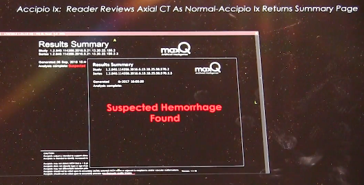
Ajay Choudhri, M.D., chairman of radiology, Capital Health, Hopewell, N.J., explains his center's experience using an artificial intelligence (AI) application to help auto detect intracranial hemorrhage. There are several AI stroke auto detection apps now available with FDA clearance or in development that were shown at the Radiological Society Of North America (RSNA) 2019 meeting. These are being adopted by hospitals and multi-center radiology practices the U.S. to flag suspected cases of ischemic stroke or brain bleeds for immediate reads. 

MaxQ AI Accipio hemorrhagic stroke detection software. MaxQ.AIThe software also offers a second set of eyes for more difficult to detect cases. Quickly determining is a stroke is ischemic or hemorrhagic is critical to the path of treatment. If caught early enough, TPA can be injected into patients to clear clots causing an ischemic stroke, but can cause massive brain damage or death if injected into a patient with a brain bleed. At advanced neuro-interventional centers, quickly determining the type of stroke is needed to know if they need to revascularize a patient or manage a hemorrhage. 


Related Content:

How Artificial Intelligence Can Predict and Detect Stroke

MaxQ AI's Intracranial Hemorrhage Software to be Integrated on Philips CT Systems

Find more news and video from RSNA

Advanced Visualization | December 30, 2019

This is a hologram of a fracture from a computed tomography (CT) scan displayed by the start up company Voxon at the 2019 Radiological Society Of North America (RSNA) meeting. The technology uses a half millimeter thick glass plate that pulses up and down very rapidly while projecting 4,000 images per second. It can display standard DICOM radiology files or STL files used for 3-D printing.

There were at least four vendors showing holographic screens to display advanced visualization 3-D renderings of anatomy from medical imaging. All four of these screens could be viewed in true 3-D using normal vision without the need for special glasses or a virtual reality visor.

The images in this example flickers because of the different frame rates of the system and the iPhone used to film it, but the actual images appears much more stable.

This technology was also included in the VIDEO: Editors Choice of the Most Innovative New Radiology Technology at RSNA 2019

Photo Gallery of New Imaging Technologies at RSNA 2019

Find more news and video from RSNA


Radiation Dose Management | December 19, 2019

Mahadevappa Mahesh, Ph.D., chief of medical physicist and professor of radiology and medical physics, Johns Hopkins University, Baltimore, treasurer of the American Association of Physicists in Medicine (AAPM),a board member of the American College of Radiology (ACR), presented a late-breaking study on how medical imaging radiation dose has started to drop over the past decade. He is the co-chair of the National Council on Radiation Protection and Measures Report (NCRP), and presented the most recent NCRP data analysis at the 2019 Radiological Society of North America (RSNA) meeting.

The new NCRP 184 report covers the period between 2006 and 2016, the period of the most current CMS data. It shows a decrease of about 20 percent in the radiation dose the U.S. population receives from medical imaging, compared to the NCRP 160 that covered the period of up to 2006.

Key findings of the study include:

   • CT dose dropped about 6 percent, despite a 20 percent increase CT scans since 2006;

   • Drop of more than 50 percent for nuclear imaging scans, mainly due to fewer procedures being performed;

   • A 15-20 percent decrease across X-ray imaging modalities.

Mahesh says this shows the impact of using "as low as reasonably achievable" (ALARA) principals, new dose guidelines outlined jointly by numerous medical societies, and dose reduction initiatives like Image Wisely, Image Gently, and the American College of Radiology (ACR) Dose Index Registry.

He said there was growing concern a decade ago when the last council report was published, which showed a steep increase in radiation dose. This was mainly due to a rapid increase in the use of computed tomography (CT) and other types of X-ray based and nuclear radiotracer medical imaging. This prompted the ACR to create the Image Wisely program and push for the use of more thoughtful imaging doses based on patient size, using the "as low as reasonably achievable” (ALARA) principle. While CT dose was lowered, he said the biggest decline over all was in nuclear imaging.


Related Medical Imaging Radiation Dose Resources:

VIDEO: Radiation Dose Monitoring in Medical Imaging — an interview with Mahadevappa Mahesh, Ph.D.

The Basics of Radiation Dose Monitoring in Medical Imaging

How to Understand and Communicate Radiation Risk — Image Wisely

Radiation in Medicine: Medical Imaging Procedures

FDA White Paper: Initiative to Reduce Unnecessary Radiation Exposure from Medical Imaging

Radiation Dose in X-Ray and CT Exams

Radiation Dose from Medical Imaging: A Primer for Emergency Physicians

Radiation risk from medical imaging

FDA: Medical X-ray Imaging


Find RSNA news and other videos


Ultrasound Transesophageal echo (TEE) | December 19, 2019

This is an example of an augmented reality (AR) training system for transesophageal echo (TEE) created by the simulation company CAE. Rather than just looking at an overhead screen, this system allows the user to use a HoloLens visor to see the impact their probe manipulation has on the cardiac ultrasound imaging and better shows the orientation of the ultrasound probe, the 2-D ultrasound image slice and the relation to the anatomy. It was displayed at the 2019 Radiological Society Of North America (RSNA) meeting.

Read more about this technology.

Find more technology news and video from the RSNA meeting

RSNA | December 18, 2019

ITN Editor Dave Fornell and ITN Consulting Editor Greg Freiherr offer a post-game report on the trends and technologies they saw on the expo floor of 2019 Radiological Society of North America (RSNA) annual meeting. This includes artificial intelligence (AI), augmented reality, holographic imaging, cybersecurity and advances in digital radiography (DR) with a glassless detector plate, X-ray tomosynthesis, dual-energy X-ray and dynamic DR imaging. 

VIDEO: Editors Choice of the Most Innovative New Radiology Technology at RSNA 2019

Photo Gallery of New Imaging Technologies at RSNA 2019

Find RSNA news and other videos


Proton Therapy | December 16, 2019

Join Chris Toth, president of Varian’s Oncology Systems business, for a peek at the history of machine learning/AI in radiation oncology, plus other highlights in 2019:

  • Ethos therapy: the world’s first AI-powered adaptive radiotherapy.
  • Noona cloud-based application for capturing patient-reported outcomes.
  • Varian’s multi-room configuration for ProBeam 360 proton therapy.
  • The promise of FLASH, an ultra-high-speed treatment that is in pre-clinical testing, and represents an exciting and potentially promising new direction in the treatment of cancer. 
Artificial Intelligence | October 22, 2019

David Sjostrom, Ph.D., deputy chief physicist, Herlev Hospital, Department of Oncology, Division of Radiotherapy, Herlev, Denmark, shares the first clinical experience treating cancer patients with the Varian Ethos radiation therapy system. He spoke to ITN at ASTRO 2019, where he presented information on the first 5 patients in the world being treated with this new technology. It uses artificial intelligence to take the onboard cone beam CT scans to automatically create an adaptive plan for any changes in patient weight loss, bladder volume, or change in tumor size. The plan can be available in minutes while the patient is on the table. It enables sparing of more healthy tissue and makes adaptive therapy much easier to use. 


Treatment Planning | August 21, 2019

This is an example of the Mirada DLCExpert deep learning software that automatically identifies organs, segments and auto-contours them as the first step in creating radiation oncology treatment plans. This example of a segmented prostate computed tomography (CT) scan being used to plan radiotherapy was created without any human intervention. It was demonstrated at the American Association of Physicists in Medicine (AAPM) 2019 meeting. 

This example shows OAR Space hydrogel (outlined in blue) injected to create space between the prostate and the rectum to prevent damage to that radiation sensitive structure. The gel is hard to identify on the CT scan because it looks like part of the rectum or prostate. But the softwares AI has been trained to identify it when present.

The DLCExpert software was cleared by the FDA in July 2018 and was first shown at ASTRO 2018. It automatically identifies anatomical structures and contours them to save staff time. The files created by the software are vendor neutral and can be imported into any vendor’s treatment planning system. Read more about this software. 

Find more news and video from AAPM

Radiology Business | August 02, 2019

Association for Medical Imaging Management (AHRA) President Chris Tomlinson, CRA, FAHRA, and President-elect Jacqui Rose, CRA, FAHRA, discuss some of the most important clinical topics at the 2019 AHRA Annual Meeting and how the association plans to help its members embrace technological change in the coming years. Among the main focuses at the meeting were clinical decision support (CDS)artificial intelligence (AI) and the use of data analytics to improve equipment and personnel performance. 

Watch the VIDEO: Assessing Cardiovascular Risk in Ultra-endurance Athletes, an interview with Colorado State University graduate research assistant Nate Bachman at AHRA 2019.

Artificial Intelligence | July 22, 2019

Leigh Conroy, Ph.D., physics resident, University Health Network, Princess Margaret Cancer Center, Toronto, Canada, explains how her center is using machine learning to automate treatment plans. The center is one of the first to use the RayStation machine learning treatment planning system for radiation oncology. She spoke at the American Association of Physicists in Medicine (AAPM) 2019 meeting. 

Learn more about how this technology works in the VIDEO: Editor's Choice of the Most Innovative Technologies at AAPM 2018.

Find more news and videos from AAPM.


Artificial Intelligence | July 12, 2019

Khan Siddiqui, M.D., founder and CEO of HOPPR, discusses the economic advantages and costs presented by artificial intelligence (AI) applications in radiology, as well as potential strategies for healthcare providers looking to add AI to their armamentarium, at the 2019 Society of Imaging Informatics in Medicine (SIIM) annual meeting.

Digital Pathology | July 11, 2019

Toby Cornish, M.D., Ph.D., associate professor and medical director of informatics at the University of Colorado School of Medicine, explains how the subspecialty of digital pathology has evolved in recent years, the benefits of integrating pathology and radiology, and how artificial intelligence (AI) may smooth the transition, at the 2019 Society of Imaging Informatics in Medicine (SIIM) annual meeting. 

Subscribe Now