Sectra provides industry-leading enterprise image management solutions comprising PACS for radiology, cardiology, and pathology, VNA and Cross Enterprise Workflow. Through 25 years of innovation and 1,700 installations, our experience in radiology has paved the way to deliver enterprise solutions that consolidate image handling and maintain workflow efficiency in the most image intense departments.
itnTV
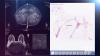
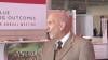
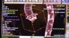
VIDEO: One on One with Amy K. Patel, MD, American Association for Women in Radiology Immediate Past President
Breast Imaging | April 15, 2024
Don't miss ITN's latest "One on One" video interview with AAWR Past President and American College of Radiology (ACR) RAN and RADPAC Chair, Amy K. Patel, MD, discussing advocacy initiatives and innovations in artificial intelligence (AI) for breast imaging.
Dr. Patel is a breast imaging trailblazer and radiology advocacy leader. In this video, learn how radiologists can support key initiatives, ways AI is improving patient care, and more.
Related content:
Technology Report: Artificial Intelligence in Radiology 2021
VIDEO: Integrating Artificial Intelligence Into Radiologists Workflow
Conference Coverage
Ultrasound Imaging | January 10, 2017

The next generation of ZONE Sonography Technology (ZST) has arrived and its living technology continues to evolve. Leveraging ZONARE’s revolutionary ZST and Mindray’s rich repertoire of workflow and user interface features, the Resona 7 is poised to become the new industry leader in premium ultrasound imaging platforms. The Resona 7 presents crystal clear B-mode imaging capabilities with unrivaled detail resolution and image uniformity across all radiology applications. Ultrasensitive Doppler modes and high-speed digital signal processing permit accurate display of hemodynamic states from skin line to depths up to 40 cm without compromising frame rate. An intuitive, customizable gesture-powered touchscreen enables logical and efficient workflow and enhanced user experience.
At its core, ZST provides unique imaging advances for the Resona 7 such as Advanced Acoustic Acquisition which renders superb imaging by using large zones to acquire up to 90 percent more acoustic data per frame and at speeds of 10 times faster than conventional technology. Dynamic Pixel Focusing creates a perfectly focused image every pixel, every frame, in every patient and in every application. Sound Speed Compensation enables a one button touch that automatically calculates the true speed of sound in a specific soft tissue and recalibrates the imaging system to optimize spatial and contrast resolution.
Finally, ZST provides Total Recall Imaging which is powerful software that allows manipulation of raw acoustic data from archived and cine images (clips) permitting a broad range of post-processing functions. This eliminates the need for repeat scanning which, in turn, aids in increasing patient throughput.
ZST is a constantly evolving software-based “living technology.” It is Mindray’s approach to providing customers with easily upgradeable ultrasound enhancements. These upgrades secure product investment protection by ensuring that ZST systems remain at the cutting-edge of imaging performance excellence throughout the system’s entire life cycle.
In summary, coupling premium imaging with advanced workflow features and user-directed ergonomic design, the Resona 7 advances premium level ultrasound imaging into the next generation.
Breast Density | January 09, 2017

Monica Saini, M.D., consultant medical director — ABUS at GE Healthcare, discusses the necessity for personalized breast care, and how Automated Breast Ultrasound (ABUS) helps meet the challenges of screening and diagnostic imaging of dense breast tissue. To learn more about Automated Breast Ultrasound, please visit gehealthcare.com/inveniaabus.
Flat Panel Displays | January 08, 2017

EIZO is proud to be entering into its 45th year of expertise, meeting hospital demands worldwide in over 80 countries. This year we are excited to unveil a few never-seen before, brand new products.
Our newest release is the RadiForce RX660, a 30-inch 6 megapixel monitor ideal for multi-modality applications. With this monitor we’re introducing the new “Work-and-Flow” that benefits radiologists today in creating an efficient and cleaner workspace.
With the Work-and-Flow, you have access to two great features:
The “Hide-and-Seek” function enables users to easily hide the Picture-in-Picture window eliminating the need for an extra monitor while still being able to access reports, patient charts, and other information.
In another feature called the “Switch-and-Go”, users can move across two workstations.
The RX660 uses the DisplayPort 1.2 Daisy Chain Connectivity for a tangle-free, easy, single cable management – this means eliminating excess wires.
Also new this year, is the RadiForce GX550, a 21.3-inch 5 megapixel, FDA approved-monitor for viewing detailed digital breast tomosynthesis and mammography images. Like the RX660, this monitor also features EIZO’s ergonomic design features.
In our CuratOR surgical solutions area, we are featuring two new products that complement our industry leading operating room video management system that allows quick access to multiple image sources and flexible arrangements across different monitors. Different workflow scenarios can be pre-defined and recalled on demand with the simple touch of a touchscreen.
Our EIZ1000 mobile large monitor tower is a turnkey large monitor mounting alternative to costly ceiling suspensions. Its sleek design and easy maneuverability enables use in multiple surgical suites. The EIZ1000 can be installed with little to no downtime, and is the optimum solution for hospitals that do not have the infrastructure to support complex ceiling suspension.
We are also expanding our OR portfolio with the release our new line of CuratOR surgical panels. These digital viewing systems consist of one or more integrated monitors – as well as IT and video management components that function as the central console in the operating room. Integrated into the hospital’s IT structure, it is ideal for work performed by operating room or nursing staff.
Finally, we are showing our CuratOR SP2-24-49 HIS/PACS configuration for viewing images and documentation. This configuration contains a 24-inch touchscreen and a 49-inch monitor forming a space-saving combination of HIS and PACS station. Different applications are covered by just one device. The polished and sealed design allows for easy cleaning.
Radiation Oncology | January 05, 2017

Join Chris Toth, president, Oncology Systems Americas, for a look at Varian’s new product introductions and major initiatives highlighted at ASTRO this year:
- The 360 Oncology care management platform, the first software system designed to integrate and coordinate integrates relevant health information so cancer patients and their care teams can collaborate on the best care. 360 Oncology brings together in a single platform, radiation, medical and surgical oncology, social services, primary care physicians, as well as the patient, to facilitate true collaborative and coordinated care.
- HyperArc high definition radiotherapy, technology that unlocks the potential of using highly non-coplanar treatment strategies to usher in a new era of precision. HyperArc is designed to automate and simplify sophisticated treatments such as stereotactic radiosurgery (SRS), and make them available to more cancer patients around the world.
- Varian’s cyber-security initiative, which is transforming the company’s software platforms to help maximize the security of patient information, maintain the integrity of treatment delivery, and enhance clinical uptime by helping defend against cyber-attacks.
Radiology Business | December 23, 2016
A discussion with Andy Colbert, managing director and founding member of Ziegler’s Healthcare Investment Banking practice, on the reasons for and strategy involved in the business trend of radiology practice consolidation. He spoke to ITN at RSNA 2016. Read the blog “Risk Abatement May Determine the Future of Radiology,” and the article “Opportunities for Growth in a Competitive Radiology Climate.”
Radiology Business | December 23, 2016
Kim Garriott, principal consultant for Logicalis Healthcare Solutions, explains the concept of value-based imaging and how it fits into healthcare reforms at RSNA 2016. Watch the related VIDEO "Value–based Imaging,” an interview with Daniel Berman, M.D., FACC, chief of Cardiac Imaging and Nuclear Cardiology, professor of imaging, Cedars-Sinai Medical Center.
Quality Assurance (QA) | December 20, 2016

Learn more about myQA, IBA’s unique platform that connects QA applications, people, and know-how through a central database and the Cloud. It offers full support throughout all of your QA, and enables you access to the different software modules and all of your data from one intuitive interface – anywhere and anytime.
RSNA 2016 | December 19, 2016
ITN and DAIC Editor Dave Fornell takes a tour of some of the most innovative new technologies being displayed on the expo floor at the Radiological Society of North America (RSNA) 2016 meeting. For key take away trends at RSNA, watch the video "Key Trends, New Technology at RSNA 2016."
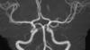
CT Angiography (CTA) | December 19, 2016
A discussion with Simon Dixon, M.D., MBChB, on the use of fractional flow reserve-computed tomography (FFR-CT) to evaluate chest pain patients in the emergency department. He is chairman of the Department of Cardiovascular Medicine at Beaumont Health System and a professor of Medicine at the Oakland University William Beaumont School of Medicine. He discussed the first year of experience with FFR-CT at Beaumont Hospital in Royal Oak, Mich., during the Transcatheter Cardiovascular Therapeutics (TCT) 2016 annual meeting. Read the article “Clinical Applications of FFR-CT.”
RSNA 2016 | December 16, 2016

A post-game roundup by ITN Contributing Editor Greg Freiherr and ITN Editor Dave Fornell on the trends and new tech seen on the show floor at the Radiological Society of North America (RSNA) 2016 meeting.
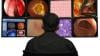
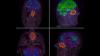
Artificial Intelligence | December 16, 2016
At RSNA 2016, the key buzzwords were “deep learning,” “machine learning” and “artificial intelligence.” Vendors and major academic centers are developing a wide array of artificial intelligence neural networks to aid radiologists in clinical diagnosis and clinical decision support. In the future, AI may also be able to help train radiologists on both normal and abnormal presentations of various organs and body systems so as to more easily identify related disease states and conditions. The following video offers two examples of how the IBM Watson system examines imaging studies.
The first case seen here demonstrates how Watson can arrive at a differential diagnosis of an aortic dissection by analyzing an abdominal computed tomography (CT) scan. The second case involves the discovery of a fibroadenoma of the breast from Watson’s analysis of a mammogram.
Watson first analyzes the text of the radiology report, identifying and pulling out key words or phrases that may indicate the potential diagnosis. It then examines the CT scan to locate relevant visible anatomic structures such as the heart, aorta and pulmonary artery. Each structure is examined for anomalies, which identifies a possible aortic dissection; the dissection is displayed within the context of the entire 3-D CT scan. Finally, Watson applies its existing clinical knowledge to the findings from the CT scan and the radiology report, establishing pathways to numerous possible conclusions until arriving at the right one.
See examples of real products using AI at RSNA 2017 in the VIDEO "Examples of How Artificial Intelligence Will Improve Medical Imaging." ITN also created an in-depth VIDEO: Technology Report — Artificial Intelligence at RSNA 2017, with interviews with numerous AI vendors.
Watch the VIDEO: “Development of Artificial Intelligence to Aid Radiology,” an interview with Mark Michalski, M.D., director of the Center for Clinical Data Science at Massachusetts General Hospital, explaining the basis of artificial intelligence in radiology.
Radiology Imaging | December 14, 2016
Patricia Oliveira-Szejnfeld, M.D., and Fernanda Tovar-Moll, M.D., Ph.D., explain what radiologists should be looking for to aid early diagnosis of Zika virus. They were among the key investigators for the first large-scale, multimodality assessment of the Zika in Brazil, the epicenter of the 2016 Zika outbreak. They spoke to ITN at RSNA 2016. Read the article “Imaging Zika Virus - Radiologic Assessment and Tracking in Prenatal Development.”
Radiation Dose Management | December 14, 2016

Mahadevappa Mahesh, MS, Ph.D., chief physicist and professor of radiology and radiological science at Johns Hopkins Hospital, explains the basics of medical imaging dose monitoring technologies. This includes monitoring and recording software meet new Joint Commission requirements, state dose laws and to improve patient safety regarding X-ray radiation exposure. Read the article “The Role of Dose Tracking Systems in Radiation Safety Programs.”
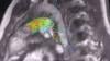
Neuro Imaging | December 12, 2016
Max Wintermark, M.D., professor of radiology and chief of neuroradiology, Stanford Hospital and Clinics, explains trends and recent advances in brain imaging at Radiological Society of North America (RSNA) 2016 meeting. He summerizes the lastest imaging technologies and hot topics in neoradiology in RSNA sessions. Wintermark has specific interest and expertise in stroke, traumatic brain injury, epilepsy, movement disorders and psychiatric disorders.
Magnetic Resonance Imaging (MRI) | December 12, 2016
Emanuel Kanal, M.D., director of MRI services and professor of radiology and neuroradiology at the University of Pittsburgh Medical Center, explains what is known about MRI contrast retention in the brain and other MRI safety concerns. He spoke to ITN at RSNA 2016.
Gadolinium-based contrast agents have been used for diagnosis and treatment guidance in more than 100 million patients worldwide over the past 25 years. These agents enhance the quality of MR images by altering the magnetic properties of nearby water molecules in the body. By improving the visibility of specific organs, blood vessels or tissues, contrast agents help physicians diagnose and treat a wide variety of medical conditions. On its own, gadolinium can be toxic. Therefore, when used in contrast agents, gadolinium is bonded with a molecule called a chelating agent, which controls the distribution of gadolinium within the body.
Read the article "Gadolinium May Remain in Brain after Contrast MRI."
Angiography | December 08, 2016
Tom Kloetzly, sales and marketing VP for Shimadzu Medical Systems USA, explains the evolution of Shimadzu Corporation since its founding 142 years ago. Kloetzly focuses on the Trinias Interventional X-ray lineshown at RSNA 2016. Kloetzly states, “A key feature of Trinias, is the ability to image from fingertip to fingertip during a Transradial approachwhich makes for much shorter hospital stay with the patient up and moving almost immediately after the procedure.Features Like RSM-DSA, a type of motion correction subtraction, eliminates patient movement during acquisition while STENTVIEW, is an enhanced visualization during stent placement in real-time.”
Artificial Intelligence | December 08, 2016
Mark Michalski, M.D., director of the Center for Clinical Data Science at Massachusetts General Hospital, explains the basis of the utilization of artificial intelligence (aka deep learning and machine learning) in radiology. He also explains where things are at in development of these neuro networks at RSNA 2016. Watch the VIDEO “Examples of Artificial Intelligence in Medical Imaging Diagnostics.”
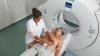
Ultrasound Imaging | December 07, 2016
Sabrina Newell, MS, RCS, clinical analyst at healthcare consulting firm MD Buyline, explains some of the trends and new technology in ultrasound at RSNA 2016. For more information about cardiac ultrasound advances, watch the video "Trends and Advances in Echocardiography at ASE 2016."
Radiation Therapy | November 18, 2016

Accuray recently unveiled the company’s newest innovation, the Radixact Treatment Delivery System, a uniquely smart, fully integrated treatment delivery, treatment planning and data management system. Hear from the Accuray team about this new, next-generation TomoTherapy platform, as well as how recent innovations to the CyberKnife platform reduce treatment time while providing leading-edge motion tracking and real-time beam adjustment. Discover how Accuray systems can help clinicians deliver precise tumor treatments with confidence.
Radiation Oncology | October 07, 2016

Rachael Bennett, clinical analyst for MD Buyline, discusses trends in radiation oncology at ASTRO 2016.
Radiation Oncology | October 07, 2016
ASTRO Chairman of the Board David C. Beyer, MD, FASTRO, discusses the current trends in radiation oncology, key trends presented at this year's conference, and achieving value, and opportunities to improve value, in the treatment of prostate cancer.
Digital Radiography (DR) | October 05, 2016

Contributing Editor Greg Freiherr offers an overview of digital radiography (DR) advances at the Association for Medical Imaging Management (AHRA) 2016 meeting. Read the article “The Coming Push for DR.” Watch a technology report sidebar video on new DR Systems technology.
Digital Radiography (DR) | October 05, 2016

Agfa highlights how its digital radiography (DR) systems capture analytics data to help improve management of the radiology department, show ROI on DR investments, and explains how its image processing software works. Read the article “The Coming Push for DR.” Watch the video “Technology Report: DR Systems.”
Enterprise Imaging | August 16, 2016

Interview with Jef Williams, managing partner, Paragon Consulting Partners, on the essential elements of building an enterprise imaging strategy, at the 2016 Association for Medical Imaging Management (AHRA) annual meeting in Nashville, Tenn. For more information on enterprise imaging technology, watch the video “Enterprise Imaging - RSNA 2015 Technology Report.”
Radiology Business | August 16, 2016

How to turn your imaging center into a Swiss army knife to maximize ROI was a key radiology business session at the 2016 Association for Medical Imaging Management (AHRA) annual meeting in Nashville, Tenn. Robert Junk, AIA, and Tobias Gilk, M.Arch, of RAD-Planning, explain how radiology departments can assess their business model to make the most of their capital investments. Read the related article that include Junk and Gilk "Rethinking the Radiology Business Model."
Breast Imaging | August 16, 2016

Interview with Gerald Kolb, JD, president of The Breast Group in Bend, Ore. Kolb spoke at the 2016 AHRA meeting in Nashville about the challenges of multimodality breast screening, the need for more personalized screening programs and the impact of recent screening recommendations. Read about the lastest advances in breast imaging.
Analytics Software | August 16, 2016

Interview with Kent Hutson, M.D., CPE, of Radiology Alliance in Nashville, Tenn., on the principles of data mining and how they can be used in radiology, at the 2016 Association for Medical Imaging Management (AHRA) annual meeting. For more information, read the article "Analytics: The Next Big Health IT Undertaking."
Quality Assurance (QA) | August 15, 2016

Patient QA Efficiency with Dolphin presented at AAPM 2016 IBA booth. Dolphin Online Ready is released and in clinical use for pre-treatment QA.
Radiation Oncology | August 12, 2016
AAPM President Bruce Curran, MEng, FAAPM, FACMP, FACR, discusses the key topics at the 2016 meeting of the American Association of Physicists in Medicine. Topics included the advancement of radiomics in medical imaging for quantification for evidence-based clinical decision making in cancer, and improving how quality assurance is done in radiation therapy.
Enterprise Imaging | July 21, 2016
The performance, feature and scalability gap between today’s enterprise viewers cannot be understated. Nearly all viewers provide image access from the EHR, but can your viewer also provide diagnostic interpretation at massive scale, with sophisticated interoperability to replace your PACS? itnTV captured Visage 7 in action at SIIM 2016, and if you watch closely, you’ll see Visage 7 explain: Fast. Powerful. Enterprise Imaging.
Information Technology | July 19, 2016

At SIIM 2016, itnTV caught up with opening keynote speaker and SIIM treasurer Rasu B. Shrestha, M.D., MBA, Chief Innovation Officer, University of Pittsburgh Medical Center and Executive Vice President, UPMC Enterprises, to discuss the dynamic changes that imaging is facing today.
Information Technology | July 19, 2016

New this year is the SIIM Innovation Challenge, which offered a prize of $10,000 to the winning team to help support innovation exploration and development to raise awareness and engagement in innovation efforts that will help shape the present and future of imaging informatics. Co-chairs Ram Chadalavada, M.D., MS, CIIP and Adam Kaye, M.D., MBA, CIIP, discussed some of the innovative ideas presented at this year’s challenge, and future plans, with itnTV.
Artificial Intelligence | July 19, 2016

Eliot L. Siegel, M.D., Dwyer Lecturer; Closing Keynote Speaker, Vice Chair of Radiology at the University of Maryland and the Chief of Radiology for VA Maryland Healthcare System, talks about the current state of the industry in computer-aided detection and diagnosis at SIIM 2016. Watch the VIDEO “Development of Artificial Intelligence to Aid Radiology,” an interview with Mark Michalski, M.D., director of the Center for Clinical Data Science at Massachusetts General Hospital, explaining the basis of artificial intelligence in radiology. Watch the VIDEO “Examples of Artificial Intelligence in Medical Imaging Diagnostics.”
Information Technology | July 19, 2016

2016 marks the third year for the SIIM Hackathon. itnTV sat down with Marc D. Kohli, M.D., Hackathon Committee-Co-Chair, SIIM Board of Directors and director of clinical informatics at UCSF to discuss some of its new objectives and future plans.
Information Technology | July 19, 2016

At SIIM 2016, Paul G. Nagy, Ph.D., CIIP, FSIIM, discussed the main challenges to adaptive change in healthcare IT, and the adaptive leadership skills necessary to complement technological changes in a clinical setting. He also discussed his vision for SIIM as he assumes leadership as incoming chair.
Computed Tomography (CT) | July 08, 2016

Interview with Claudio Smuclovisky, M.D., FACC, FSCCT, director of South Florida Imaging Cardiovascular Institute, Holy Cross Hospital, at the Society of Cardiovascular Computed Tomography (SCCT) 2016 annual meeting. Smuclovisky explains what imaging departments need to know about when purchasing the newest generation of CT scanners. He explains there is more to scanners than slices, offering information beyond the hype over 64-, 128-, 256-, 320-, and 640-slice CT scanners. For more information, read "Costs vs. Benefits: Comparing 64-Slice to 256, 320-Slice CT."
Computed Tomography (CT) | July 08, 2016
Interview with Patricia Dickson, LRT (CT), assistant director, diagnostic and outpatient services, Capital Cardiology Associates, Albany, N.Y., at the Society of Cardiovascular Computed Tomography (SCCT) 2016 annual meeting. She explains what technologists need to know when prepping patients and imaging during cardiac CT exams. For trends in cardiac CT, watch the VIDEO "The Future of Cardiac CT in the Next Decade."
Computed Tomography (CT) | July 08, 2016
An interview with Jonathan Leipsic, M.D., FSCCT, chairman of the department of radiology, St. Paul’s Hospital, Vancouver, Canada, at the Society of Cardiovascular Computed Tomography (SCCT) 2016 meeting. Leipsic is heavily involved with the procedural planning and anatomical assessments for TAVR and clinical trials for new transcatheter mitral valves and annulus repairs.
CT Angiography (CTA) | July 07, 2016
DAIC/ITN editor Dave Fornell shows some of the most innovative new cardiac CT and angiography technologies from sessions and the expo floor at the Society of Cardiovascular Computed Tomography (SCCT) 2016 annual meeting.
© Copyright Wainscot Media. All Rights Reserved.
Subscribe Now