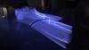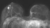
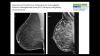
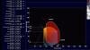
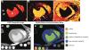
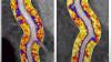
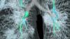
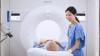
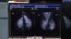
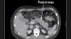
Professor Christiane Kuhl, M.D., director of radiology, University Hospital Aachen, Germany, explains how breast magnetic resonance imaging (MRI) can be used to clearly identify breast cancers in women with dense breast tissue. In women with dense breasts, it can be very difficult to detect many cancers on standard mammograms because the cancers and dense tissue both appear white. MRI can help clearly define tumors and identify which nodules are cancer and which are benign, which can help greatly reduce the need for biopsies.
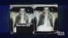
Kuhl is an expert in breast imaging and breast MRI. She helped develop an a shortened MRI protocol that allows breast MR images to be created in 3 minutes or less, rather than standard protocols that can take up to 30 minutes. In the interview she shows patient case examples of standard mammograms and the MRI supplemental imaging for the same patient to show the hidden tumors.
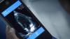
She also explains the differences between standard 2-d mammography, the current standard of care, and the newer 3-D mammogram tomosythnesis technology, breast ultrasound and breast MRI technologies.
Other video interviews with Dr. Kuhl:
VIDEO: Explaining Dense Breasts
VIDEO: The Impact of COVID-19 on Breast Imaging
Related Breast MRI Content:
Abbreviated MRI Outperforms 3-D Mammograms at Finding Cancer in Dense Breasts
VIDEO: Explaining Dense Breasts — Interview with Christiane Kuhl, M.D.
VIDEO: Use of Breast MRI Improved Cancer Detection in Dense Breasts in Dutch Study — Interview with Gillian Newstead, M.D.
Technologies to Watch in Breast Imaging
Screening MRI Detects BI-RADS 3 Breast Cancer in High-risk Patients
Rapid Breast MRI Screening Improves Cancer Detection in Dense Breasts


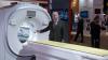


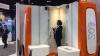
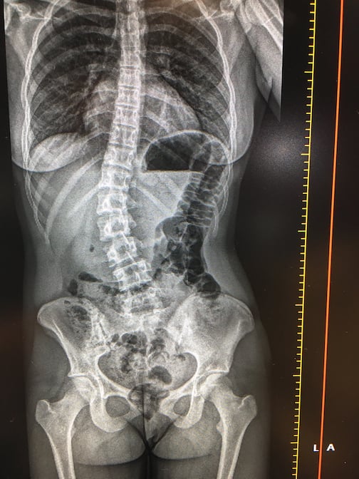 This is a demo of the EOS orthopedic X-ray imaging system at the recent 2019