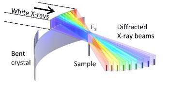
How the bent crystal changes the direction of the X-rays. Image courtesy of Tohoku University
May 15, 2020 — Many will undergo a computed tomography (CT) scan at some point in their lifetime — being slid in and out of a tunnel as a large machine rotates around. X-ray computed tomography, better known by its acronym CT, is a widely used method of obtaining cross-sectional images of objects.
Now a research team — led by Tohoku University Professor, Wataru Yashiro — has developed a new method using intense synchrotron radiation that produces higher quality images within milliseconds.
High-speed, high-resolution X-ray CT is currently possible using intense synchrotron radiation. However, this requires samples to be rotated at high speed to obtain images from many directions. This would make CT scans more akin to a rollercoaster ride!
Extreme rotation also makes controlling the temperature or atmosphere of the sample impossible.
Nevertheless, the research team solved this conundrum by creating an optical system that splits single synchrotron X-ray beams into many. These beams then shine onto the sample from different directions at the same time; thus, negating the need to rotate the sample.
This "multi-beam" method is no easy task since the direction of X-rays cannot be easily changed. Unlike visible light, X-rays interact with matters weakly, making it difficult to utilize mirrors and prisms to change the path of the beams.
To overcome this, the research team used micro-fabrication techniques to create uniquely shaped crystals. These crystals were then bent in the shape of a hyperbola. By combining three rows of crystals, the multi-beam optics were able to cover an angle of ±70°.
Carrying out their experiments at the SPring-8 synchrotron radiation facility, the research team took advantage of a cutting-edge compressed-sensing algorithm that needs only a few dozen projection images for image reconstruction.
"The invention makes 3-D observations of living beings and liquid samples within milliseconds possible" exclaimed Yashiro. "It is possible application is wide-spread, from fundamental material science to life sciences to industry."
For more information: www.


 April 22, 2024


