A promising imaging technology that measures bone health at the microscopic level could help doctors detect osteoarthritis in its early stages and guide the development of new therapies, suggests preliminary research being presented at the 58th Annual Meeting of the American Association of Physicists in Medicine (AAPM).
“The technology we are developing allows us to see very fine detail in the mesh-like microstructure of bone — known as trabecular bone — which currently can’t be assessed in patients,” said Wojciech B. Zbijewski, Ph.D., a medical physicist and instructor in the department of biomedical engineering at Johns Hopkins University, Baltimore. “This could help us detect bone diseases in their initial stages and help with development of new preventive therapies. For example, there is growing evidence that early stages of osteoarthritis involve changes in trabecular bone, so if we can detect such change, a patient could potentially avoid painful knee replacement surgery by getting treatment before the cartilage was irreversibly damaged.”
Osteoarthritis often occurs after a joint is injured, leading to progressive damage of the cartilage that lines and cushions the joint and causing pain and stiffness.
The technology, a special cone beam computed tomography (CBCT) system using complementary metal-oxide semiconductor (CMOS) detectors provides several advantages over standard methods: the device is portable and could be used in doctors’ offices; patients could be imaged standing (vs. lying down); and the radiation dose is lower than standard computed tomography (CT). The CMOS detector provides very high spatial resolution, allowing detection of finer details than in standard CT and therefore greatly enhancing the ability to assess trabecular bone. This imaging system also could help with early identification of osteoporosis, which causes bones to become weak and brittle and break more easily.
The researchers used CBCT/CMOS to image the hand of a human cadaver, as well as pelvic bone biopsy samples from 25 living women. They determined it provided results that correlate with micro-CT, which is the gold standard for assessing trabecular bone, but can only be used for bone samples (biopsies), not living patients whose joints are too large to be scanned using this method. They anticipate beginning the first studies of CBCT/CMOS in patients late next year.
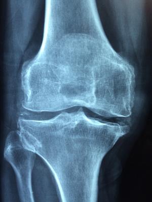
Today, joint pain often is assessed by X-ray or magnetic resonance imaging (MRI), which shows if the cartilage lining the joint has been damaged. In most cases, by the time a patient has pain, the damage to the joint is irreversible. The only permanent solution is to surgically replace that joint. Joint replacement is a common procedure: 719,000 knee replacements and 332,000 hip replacements are performed every year, according to the Centers for Disease and Prevention (CDC).
If further research supports these findings, in the future a person who injures a joint — for example by breaking a bone or tearing a ligament — could be scanned using this technology to assess the bone health immediately after the injury and during treatment. Current prevention methods include losing weight, doing exercises that don’t stress the joint (such as swimming and biking) and taking supplements such as vitamin D and calcium. This technology also could be used by researchers to guide development of treatments to prevent bone damage, said Zbijewski.
In addition to Zbijewski, collaborators on the study being presented at AAPM include: Qian Cao, Alejandro Sisniega, Michael Brehler, J. Webster Stayman, Jeffrey Siewerdsen, Eugenio Marinetto, and John Yorkston. The research was made possible through a grant from the National Institutes of Health.
For more information: www.aapm.org

 April 17, 2024