May 17, 2017 — Tumors, inflammation and circulatory disorders locally disturb the body's acid-base balance. These changes in pH value could be used for example to verify the success of cancer treatments. Up to now, however, there has been no imaging method to render such changes visible in patients. Now a team from the Technical University of Munich (TUM) has developed a pH sensor that renders pH values visible through magnetic resonance imaging (MRI) – in a non-invasive, radiation-free manner.
Four years ago, during a magnetic resonance experiment with tumor cells, TUM physicist Franz Schilling, Ph.D., found signals from a molecule that was highly sensitive towards pH changes. The molecule, which was identified as zymonic acid in subsequent investigations, could play an important role in the future of medical imaging. As a biosensor for pH values, it could provide insights into the body which had been impossible in the past.
"An appropriate pH imaging method would make it possible to visualize abnormal changes in tissue and specifically metabolic processes of tumors," explained Schilling. Areas surrounding tumors and inflammations are usually slightly more acidic than areas surrounding healthy tissue, a phenomenon possibly linked to the aggressiveness of tumors. Schilling sees further potential uses in treatment prognoses. "pH values are also interesting when it comes to evaluating the efficacy of tumor treatments. Even before a successfully treated tumor starts to shrink, its metabolism and thus the pH value of the surrounding area could change. An appropriate pH imaging method would indicate at a much earlier stage whether or not the right approach has been selected," he said
Schilling is now director of the working group for Preclinical Imaging and Medical Physics at the Clinic and Polyclinic for Nuclear Medicine in the TUM Klinikum rechts der Isar. In past years, he has joined together with colleagues from the departments of physics, chemistry and medicine to research zymonic acid as a biosensor. In the journal Nature Communications, the team describes how it can be used to reliably represent pH values in the bodies of small animals.
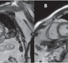
In order to make pH values visible using zymonic acid, the molecule is injected into the body and then an MRI investigation is made of the object tissue. In a strong magnetic field, radiowaves excite the nuclear spins of the zymonic acid to oscillation. The reactions of the nuclei are then recorded. This data is used to calculate frequency spectra that in turn provide information about the chemical properties of the molecular surroundings of the nuclei. Ultimately, the pH value at any examined location in the tissue can be represented based on pH-dependent molecular changes in the zymonic acid.
Zymonic acid has to be marked with carbon-13 in order to be visible in MRI images. This means that the molecules contain carbon-13 atoms (13C) instead of "normal" carbon 12 atoms. But zymonic acid marked in this manner is still not measurable; its MRI signal is too weak. "We therefore use a relatively new method, hyperpolarization," explained Stephan Düwel, physicist and first author of the study. "We use a special device to transfer the polarization of electrons to the 13C atomic nuclei using microwaves at very low temperatures, which results in an MRI signal up to 100,000 times stronger." A hot liquid is then used to quickly return the zymonic acid to room temperature.
After this, the scientists need to act quickly. The biosensor is injected intravenously into the organism, then the MRI scan has to be made immediately: It only takes 60 seconds for the signal-amplifying effect of the hyperpolarization to wear off again. "We're currently working on expanding this time window," said Düwel.
"On the one hand, we're trying to improve the MRI properties of zymonic acid with appropriate modifications to the molecule; On the other hand, we're looking for other pH-sensitive molecules," explained biochemist Christian Hundshammer, second author of the study.
Schilling and his team have succeeded in showing that their method is sensitive enough to represent medically relevant pH value changes in the organism. Using zymonic acid it is furthermore possible to specifically investigate the pH value outside of the cell membrane: With other biosensors it is often not clear whether measured changes take place inside or outside of the cell (intracellular or extracellular). This is important because the intracellular value is usually stable, while changes in metabolism have a much greater impact on the extracellular value.
In contrast to optical methods, which are limited to superficial penetration into the body because of the low transparency of tissue, there are no limitations to the depth of penetration for MRI. It has furthermore been demonstrated that zymonic acid is not toxic in the concentrations used with small animals and is also created in low concentrations as a by-product of the metabolite pyruvic acid which is present in the body.
"We believe zymonic acid is a highly promising biosensor for patient applications," said Schilling. For the time being, however, additional pre-clinical studies are planned in order to ascertain the advantages of this new imaging biomarker compared to conventional methods and to further improve the spatial resolution of pH imaging.
The research project was funded by the Collaborative Research Centre 824 (SFB824) “Imaging for Selection, Monitoring and Individualization of Cancer Therapies” led by Prof. Markus Schwaiger.
For more information: www.nature.com/ncomms

 April 17, 2024