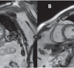
Using fast MRI, UC Davis researchers scanned left and right wrists of men and women and used the data to build computer models of the movement of wrist bones. The data could help understand wrist injuries such as carpal tunnel syndrome. Image courtesy of Brent Foster and Abhijit Chaudhari, UC Davis.
February 19, 2019 — In a just-published Journal of Biomechanics article1, the researchers proved a longtime assumption about individuals' right and left wrists, while also finding differences between wrists of males and females. These discoveries could help inform and guide future treatments.
"If someone has dysfunction of the wrist, it really impacts their quality of life," said UC Davis Biomedical Engineering Graduate Group member and article first author Brent Foster, who also was selected for a prestigious National Science Foundation Graduate Research Fellowship to create a wrist-anatomy model. Foster currently is a graduate student in the lab of Abhijit Chaudhari, Ph.D., in the UC Davis Department of Radiology, and his co-authors include orthopedic surgeons, a radiologist and medical imaging experts at UC Davis and the University of Southern California.
Foster and his team scanned both wrists of 18 individuals — nine men and nine women of varying ages — with no history of wrist injuries, disease or pain. Using innovative magnetic resonance imaging (MRI) techniques that allow 3-D views of wrists in motion, the researchers had individuals move their wrists in five ways while being scanned to track the movement of the wrist bones and joint. They then used advanced mathematical techniques to analyze the images to generate robust models of wrist motion.
"While each wrist bone had been studied individually before, our work really focuses on how wrist bones move and act together," Foster said.
The results showed that while individual's left and right wrists are similar, there were gender-based wrist differences. While wrist-based therapies had assumed that individuals' left and right wrists mirrored each other, there has been insufficient evidence to back this assumption up until now.
The researchers initially hypothesized that there would not be significant wrist differences found between the male and female volunteers. But their measurements changed their minds: "While there is literature about scaling differences between male and female wrists, we are able to examine if bone trajectories during wrist motion differ by gender," said Foster.
Wrist conditions such as carpal tunnel syndrome do disproportionately affect women, although it's not clear why, Chaudhari said.
"There have historically been several theories about what wrist bones do during motion, and some cadaveric studies to support them. Analysis performed based on some of these theories illustrates sex differences, but that based on others doesn't," he said. The new fast MRI and computed tomography (CT) scanning techniques allow researchers to study live wrist bones in motion for the first time.
Foster, Chaudhari and colleagues look forward to continuing their efforts to help people experiencing wrist pain and disorders like osteoarthritis and carpal-tunnel syndrome.
"By scanning just five basic wrist movements, we were able to explain over 91 percent of wrist variation across individuals," he said. "We're excited to use these innovative MRI and analysis methods to make a difference in managing wrist disorders."
For more information: www.journals.elsevier.com/journal-of-biomechanics
Reference

 April 04, 2024