
Greg Freiherr has reported on developments in radiology since 1983. He runs the consulting service, The Freiherr Group.
Digital PET Balances Scan Time and Resolution

Staging F18FDG PET/CT images of adenocarcinoma in the RUL (right upper lobe) of the lung illustrates the value of Vereos. The primary lesion in the right upper lobe appears in the upper row (PET image is left, CT image is right). A 3 mm synchronous primary or metastatic lesion in the RUL is apparent in the lower row. The precision afforded by Vereos' images provided the basis for the patient to undergo RUL lobectomy instead of thermal ablation of the primary lesion. (Images courtesy of Dr. Jay Kikut and UVMC)
Efficiency and effectiveness are inseparable in clinical medicine. Digital PET addresses them both. The key is the detector built into Philips Healthcare’s digital Vereos PET/CT.
Because photons generated during a PET exam are counted individually, digital detectors can record more such events per second than analog ones.
The University of Vermont Medical Center (UVMC) often leverages this on its Vereos to produce images with very high quality. Alternatively, UVMC uses Vereos to shorten scan time while still producing diagnostic quality images.
“When there is a big leap in sensitivity like you see from analog to digital, you have to look at what you are going to do with it,” said Jay Kikut, M.D., director of nuclear medicine and PET/CT at the UVMC. “For oncology patients, our decision is clear — we want to use it for improved image quality.”
This is paramount when clinicians use Vereos to make diagnoses and stage cancer patients. “Vereos provides us very accurate staging of our patients,” he said. “To have the best outcome, you have to match the treatments to the stage.” Exactly staging patients leads to a more individualized choice of therapy.
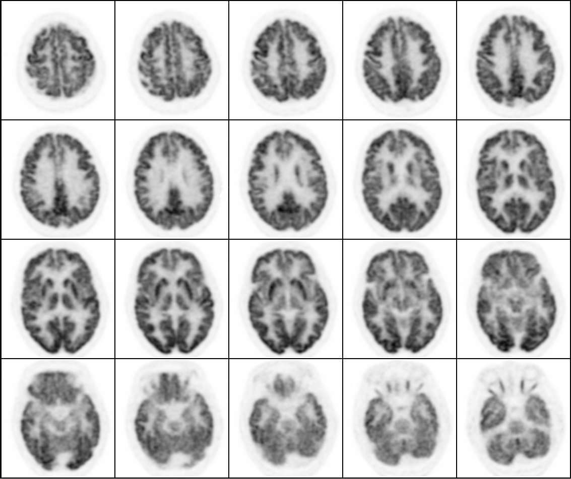
The 1 mm thick images reveal details in this PET/CT whole-brain volume acquired during a five-minute scan on the Vereos PET/CT. Images courtesy of Philips Healthcare.
Oncological applications account for about 80 percent of PET scans done at UVMC. (The remaining 20 percent of PET/CTs examine the heart or brain.)
When set to deliver maximum spatial resolution, the increased sensitivity achieved through digital PET is used to detect very small lesions. Alternatively, some patients at UVMC are best served by scans that minimize time spent inside the PET/CT bore. Such shorter exams might be chosen for children or patients who are uncomfortable in tight spaces to minimize the movement that can cause image artifacts.
Although the scans may be substantially shorter — five or even three minutes versus the 15 needed with an analog detector — Vereos’ PET acquisitions can still deliver high diagnostic quality, he said.
Development of the Digital Photon Counting (DPC) technology, which serves as Vereos’ backbone, is the latest pivotal moment in the history of PET/CT, according to Dhruv Mehta, a senior product manager at Philips Healthcare. The first occurred some 20 years ago with the hybridization of PET with CT. The second was the development of time-of-flight (introduced first-to-market by Philips Healthcare), which helps localize lesions and improves signal-to-noise. Fully digital PET with DPC technology is the latest advancement affecting this hybrid, Mehta said.
“It is really the next generation of PET/CT,” he said.
The difference between digital and analog PET is akin to the difference between projection and LCD televisions. The digital architecture of LCD TVs, he said, delivers a sharper image, higher efficiency and more dynamic range. Digital PET does much the same in molecular imaging. This linkage between image quality and dynamic range is important, Mehta said, when trying to go beyond the current FDG-based oncology applications into emerging applications in neurology and cardiology and emerging tracers.
“With a shorter acquisition, the patient is less likely to move,” he said. “The clinical benefit is that you have a better study with less motion. As a result, you can visualize lesions better.”
The other aspect of efficiency is increased throughput. In the U.S., PET patient volumes typically hover around a dozen or so patients per day. In Europe or China, however, daily throughput may exceed 30.
Mehta cautions that many factors beyond the scanner may affect throughput at a facility. Chief among them are ones related to patient management. Cutting scan time to under five minutes, however, “is a substantial improvement.”
Very relevant when making purchasing decisions, he said, is the increased precision that digital PET offers and the hedge that Vereos provides against obsolescence. At present, PET is one of the few commercial modalities with high-end systems that are still analog, he said. That, however, could change.
“We envision a time in the future when all PET/CT is digital,” Mehta said.
Editor's note: This is the third blog in a series of four on digital PET/CT. The first blog, “How Digital PET/CT Can Improve Clinical Care,” can be found here. The second blog, “Why - And How - Digital PET Is Better Than Analog,” can be found here.
Related Content:
Related Content

News | Cardiac Imaging
April 23, 2024 — CDL Nuclear Technologies, a pioneer in advanced diagnostic solutions, is proud to announce the launch ...
 April 23, 2024
April 10, 2024 — Blue Earth Therapeutics, a Bracco company and emerging leader in the development of innovative next ...
April 10, 2024 
April 5, 2024 — RLS Radiopharmacies, America’s only Joint Commission-accredited radiopharmacy network, today announced ...
April 05, 2024 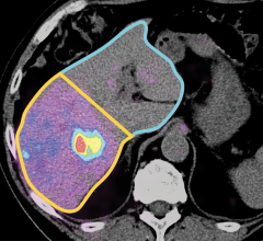
News | Radiology Business
April 2, 2024 — Less than three months after signing an agreement to acquire MIM Software Inc., GE HealthCare ...
April 02, 2024 
News | Molecular Imaging
March 29, 2024 — Magnetic resonance imaging (MRI) is a cornerstone in the landscape of medical diagnostics, celebrated ...
March 29, 2024 
February 21, 2024 — Blue Earth Therapeutics, a Bracco company and emerging leader in the development of innovative next ...
February 21, 2024 
February 14, 2024 — PanTera, the Belgian joint venture created by IBA and SCK CEN to secure large-scale production of ...
February 14, 2024 
News | Quality Assurance (QA)
February 12, 2024 — IBA, a world leader in particle accelerator technology and a world-leading provider of dosimetry and ...
February 12, 2024 
News | FDA
February 12, 2024 — Proscia, a leading provider of digital and computational pathology solutions, has received 510(k) ...
February 12, 2024 ![(A) PET images of [68Ga]Ga-DOTA-ZCAM241 uptake at baseline and 3, 7, and 12 days after injection as inflammatory arthritis developed in single representative individual mouse. Images are normalized to SUV of 0.5 for direct comparison between time points. (B) CD69 immunofluorescence Sytox (Thermo Fisher Scientific) staining of joints of representative animals during matching time points.](/sites/default/files/styles/feed_medium/public/PET%20Tracers.jpeg?itok=P5Di6MIe)
News | PET Imaging
February 9, 2024 — A novel PET imaging technique can noninvasively detect active inflammation in the body before ...
February 09, 2024 © Copyright Wainscot Media. All Rights Reserved.
Subscribe Now