January 28, 2019 — The Radiological Society of North America (RSNA) presented its seventh Alexander R. Margulis Award for Scientific Excellence to Paras Lakhani, M.D., from Thomas Jefferson University Hospital (TJUH) in Philadelphia, for the article, “Deep Learning at Chest Radiography: Automated Classification of Pulmonary Tuberculosis by Using Convolutional Neural Networks.” Lakhani was presented with the award at the 2018 RSNA annual meeting, Nov. 25-30 in Chicago.
Named for Alexander R. Margulis, M.D., a distinguished investigator and inspiring visionary in the science of radiology, this annual award recognizes the best original scientific article published in RSNA’s peer-reviewed journal Radiology.
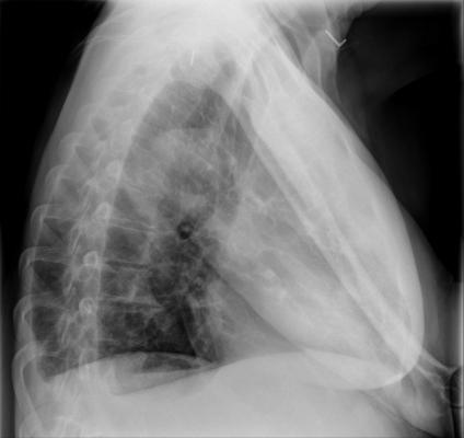
While imaging plays a pivotal role in the diagnosis and management of tuberculosis (TB), access to radiology is often limited in the developing countries where TB is most prevalent. Hoping to bridge that gap, Lakhani and colleague Baskaran Sundaram, M.D., also from TJUH, investigated the efficacy of an automated method for detecting TB on chest radiographs. Specifically, the researchers used deep learning, a type of artificial intelligence (AI) using pre-trained deep convolutional neural networks (DCNNs), to identify TB on chest X-rays. The results of the research were promising.
“We determined that deep learning with DCNNs can classify TB at chest radiography,” said Lakhani, lead author on the study. “This method means that radiography may facilitate screening and evaluation efforts in TB-prevalent areas with limited access to radiologists.”
This type of innovative research represents the future of radiology, according to Radiology editor David A. Bluemke, M.D., Ph.D.
“The authors evaluated a worldwide problem in public health — especially for areas with few radiologists,” Bluemke said. “Importantly, Drs. Lakhani and Sundaram validated their results by studying chest-X-rays from the United States, Belarus and China. This type of well-validated study is going to change the practice of radiology.”
The potential for improving detection of TB, one of the top 10 causes of death worldwide, was a strong motivator for the research, Lakhani said. In 2016, approximately 10.4 million people fell ill from TB, resulting in 1.8 million deaths, according to the World Health Organization (WHO).
“An automated solution — or proof that an automated solution could work — could change the landscape of this disease, particularly in developing countries like Sub-Saharan Africa,” Lakhani said. “A great priority of WHO is ending TB.”
For the study, Lakhani and Sundaram obtained 1,007 X-rays of patients with and without active TB, consisting of multiple chest X-ray datasets from the National Institutes of Health, the Belarus Tuberculosis Portal and TJUH. The datasets were split into training (68 percent), validation (17.1 percent) and test (14.9 percent).
The cases were used to train two different DCNN models – AlexNet and GoogLeNet – which learned from TB-positive and TB-negative X-rays. The models’ accuracy was tested on 150 cases that were excluded from the training and validation datasets. The best performing AI model was a combination of the AlexNet and GoogLeNet, with a net accuracy of 96 percent.
The two DCNN models had disagreement in 13 of the 150 test cases. For these cases, the researchers evaluated a workflow where an expert radiologist was able to interpret the images, accurately diagnosing 100 percent of the cases. This workflow, incorporating a human into the loop, had a greater net accuracy of close to 99 percent.
The DCNNs were not trained to distinguish potential mimics of pulmonary TB, such as lung cancer, bacterial pneumonia or tropical diseases, according to Lakhani.
“The goal of such algorithms is to differentiate normal from abnormal chest X-rays with respect to TB evaluation,” Lakhani said. “Those flagged as abnormal with characteristics of pulmonary TB should be followed by bacteriologic confirmation, as suggested by screening workflows presented by WHO. The goal in these workflows is cost savings, as the cost of digital radiography has substantially lowered in the past decade.”
Lakhani, who completed his fellowship training in nuclear medicine and positron emission tomography/computed tomography (PET/CT), has been a radiologist since 2011, primarily specializing in cardiac radiology at TJUH. He said, along with being a tremendous honor, the Margulis Award provides momentum for his plans to further improve the models with more training cases and other deep learning methods.
“This award was so unexpected, and I am truly honored,” Lakhani said. “Artificial intelligence is a hot area of research, and I have been focusing on this area for about two years. I don’t plan to change direction any time soon.”
While this was a retrospective study based on datasets available at the time of the study, Lakhani hopes to broaden the study by investigating the use of DCNNs in a clinical practice for evaluating TB.
“With deep learning, the more data you have, the better you do,” he said. “There is a plenty of data internationally to develop more robust algorithms, and the future is exciting for this type of research.”
For more information: www.pubs.rsna.org/journal/radiology
Reference
1. Lakhani P., Sundaram B. Deep Learning at Chest Radiography: Automated Classification of Pulmonary Tuberculosis by Using Convolutional Neural Networks. Radiology, April 24, 2017. https://doi.org/10.1148/radiol.2017162326

 April 17, 2024