Philips' ScanWise software automatically sets MRI protocols based on MR-conditional implantable devices the patients has. This is supposed to speed workflow and improve image quality.
The most recent advances in magnetic resonance imaging (MRI) technology have been on the software side, enabling faster contrast scans, greatly simplified cardiac imaging workflows, and allowing MR scans of the lungs. In addition, a few new MRI scanners have entered the market in the past year.
Watch the video “MRI Technology Report at RSNA 2015.” Contributing Editor Greg Freiherr offers an overview of MRI advances at the Radiological Society of North America (RSNA) 2015 annual meeting.
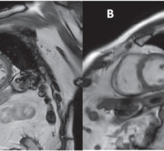
Multi-Contrast MRI Images From a Single Acquisition
In September, the U.S. Food and Drug Administration (FDA) granted market clearance for GE Healthcare’s MAGiC (MAGnetic resonance image Compilation) software, the industry’s first multi-contrast MRI technique that delivers eight contrasts in a single acquisition in a fraction of the time of conventional imaging. MAGiC is the result of a collaboration with SyntheticMR AB and gives clinicians more data than conventional scanning. This technique gives users the flexibility to manipulate MR images retrospectively, leading to significant timesavings, fewer rescans and therefore cost savings, which combined can assist the clinician in making a more decisive diagnosis. It uses an acquisition technique that allows the ability to modify image contrast after scanning has been completed, which is not possible with conventional MR. GE said MAGiC delivers more potential for the clinician to utilize changes in the image contrast to enhance their diagnosis and to reduce orders for rescans. Clinicians have the ability to generate multiple image contrasts in a single MR scan – including T1, T2, STIR, T1 FLAIR, T2 FLAIR, dual IR, phase sensitive IR and proton density weighted images of the brain in a single acquisition. To change the contrast, users simply move the cursor on the interface to change the parameters such as TR, TE and TI. MAGiC enables one scan that can do the work of many. MAGiC is available on GE’s Optima MR450, Optima MR450w, Discovery MR750, Discovery MR750w and Signa Pioneer MRI systems.
Lung MRI Now Possible
Lung MRI has been problematic since the lungs are filled with air and there is a low density of the hydrogen atoms required to create MR images. For this reason, computed tomography (CT) has traditionally been used for lung imaging. At RSNA 2015, Toshiba introduced its Ultrashort Echo Time (UTE) sequence for dedicated pulmonary MRI. Available on the Vantage Titan 3.0T MR system, UTE allows clinicians to view tissue with very short relaxation times and high susceptibility regions where signals generally disappear too quickly for accurate MR imaging.
Software Greatly Reduces MRI Scan Times
In April, the FDA approved two new Siemens MRI applications. The Simultaneous Multi-Slice application acquires MR images simultaneously as opposed to sequentially, reducing 2-D acquisition times by as much as a factor of 8. GOBrain is designed to dramatically reduce the time required for MRI examinations of the brain. Shorter scans are increasingly important at a time when brain scans account for approximately 1 out of every 4 MRI examinations, and when the number of brain MRI exams is expected to swell to 45 million worldwide this year, Siemens said. Using SMS, physicians can reduce the length of MRI brain examinations, which can vary significantly, to times compatible with the clinical routine (e.g., up to 68 percent for diffusion tensor imaging, or DTI) and bring clinical relevance to advanced neurological applications. SMS can be used in the treatment of patients who possess limited tolerance for longer scan times, including pediatric or geriatric patients. In brain surgery cases, SMS may facilitate surgical mapping and improve efficiency in the OR. Enabling clinically validated brain examinations in just five minutes, the new GOBrain application allows acquisition of clinically essential image orientations and contrasts with a single button-push. The technology is backed by Siemens’ high-channel density coils and the company’s MRI scanning software DotGO. GOBrain helps improve patient throughput and potentially reduce costs per scan. Shorter scan times, which are better tolerated by patients, can help curb lengthy and potentially expensive rescans as well as potentially reduce sedation.
The SMS application is available on Siemens’ Magnetom Aera 1.5T, Magnetom Skyra 3T, and Magnetom Prisma and Prisma Fit 3T MRI systems. The GOBrain application is featured on the Magnetom Aera and Magnetom Skyra systems.
Simplifying Cardiac MRI
Cardiac MRI has been very limited, only making up about 1 percent of all MRIs in the United States due to its complexity, long exam times and high cost. However, GE Healthcare introduced a new MRI technology at RSNA 2015 to greatly simplify cardiac MR in hopes of expanding its adoption in place of CT scans. Developed for its new Signa MRI scanners, the new ViosWorks cardiac MRI software helps automate the image sequences to perform a full 3-D chest volume scan that includes the full motion of the myocardium during the cardiac cycle, blood flow, time and fully automated quantification to create what GE calls a 7-D cardiac MRI exam. ViosWorks also speeds the imaging time from 70 minutes down to about 10 minutes using a single, free breathing exam.
Gathering a full volume dataset of a chest in motion creates a large amount of data that would normally clog the average picture archiving and communication system (PACS) and post-processing 3-D image workstation. An average cardiac MRI exam today is about 200 MB, while a ViosWorks exam is about 20 GB. So, GE has launched a new cloud computing service to help process that large amount of data quickly with remote super-computing power.
Read the article "Cardiac MRI Advances."
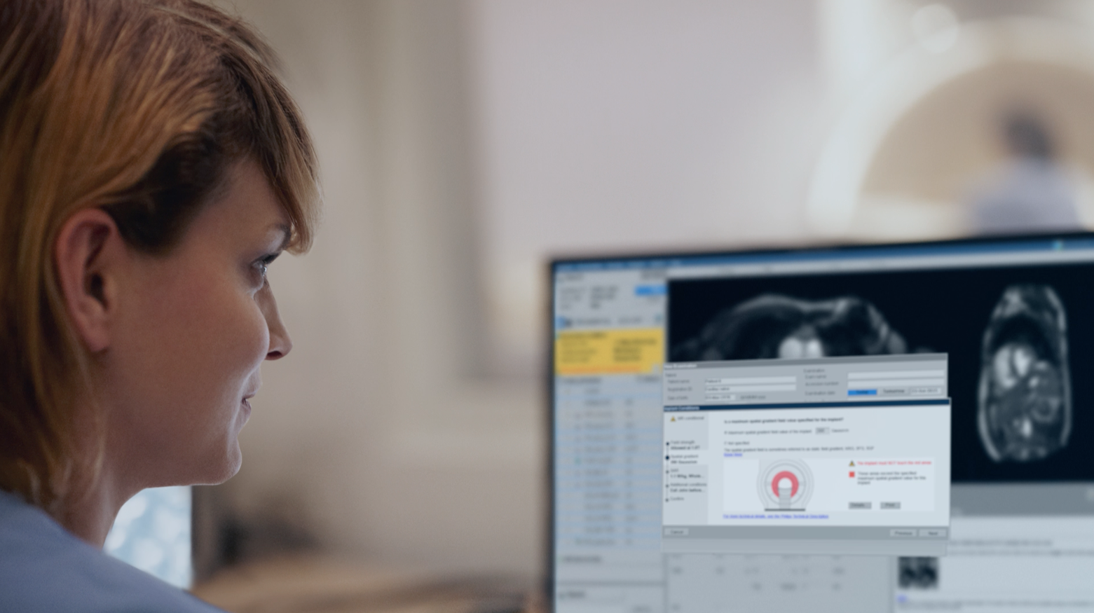
Simplifying MRI-Conditional Implant Scans
Advancements in medical implant technologies have greatly increased the number of patients with these devices in recent years, which complicated MRI scanning of these patients. MR-conditional implants include knee and hip replacements, spine implants, pacemakers and implantable cardioverter defibrillators (ICDs). More of these devices are becoming MR-conditional, which permits MRI scanning within certain parameters. These implants require adjustments in the scanner setup in order to create optimal images in the presence of these metal implants and to ensure patient safety. However, these adjustments can be time consuming and complex, causing patients with MR-conditional implants to often be denied MRI exams. At RSNA 2015, Philips unveiled the first MRI automated user interface technology designed to greatly simplify exams with patients that have MR-conditional medical implants. The new ScanWise Implant software offers a guided user interface and automatic scan parameter selection designed to support “first-time-right” imaging. The software guides operators to meet the specific criteria for each implant.
FDA Clears First 7T MRI System
In October 2017, the FDA cleared the first 7T MRI system, more than doubling the static magnetic field strength available for use in the United States. The Magnetom Terra from Siemens Healthineers is the first 7T MRI system cleared for clinical use in the United States. The first approved indications for the system are The system’s neurological and musculoskeletal (MSK). Read the article.
Hitachi Expands Apps for MRI
Last fall, Hitachi Medical Systems America released its new MRI software platform Evolution 5 for current Hitachi customers and new system orders. Evolution 5 offers nearly 40 new features and improvements implemented that continue to raise the level of imaging performance and to expand the breadth of diagnostic applications. With the integration of a new image processing algorithm (VIVID), new reconstruction task (NCC) that reduces redundant noise pickup and new optimized fast-spin echo sequence (opFSE), customers that migrate to Evolution 5 will see dramatically improved sharpness and clarity. The software also includes applications for motion compensation, a neuro suite, selective saturation for controlled MR angiography (MRA) evaluation in the abdomen, diffusion with enhanced functional analysis including kurtosis imaging (DKI), liver imaging with iron quantification, and breast imaging with breast spectroscopy for therapy monitoring.
Silent MRI Scanning
GE Healthcare has expanded its SilenScan MRI noise reduction technology to its Signa Pioneer 3T system, which features an enhanced SilentScan package to greatly reduce noise during MRI scans. SilentScan has been added for musculoskeletal (MSK) imaging and spine imaging, in addition to a complete neuro exam that also includes diffusion weighted imaging (DWI). SilentScan includes two distinct approaches to reduce acoustic noise by addressing the source, the gradient-magnet interaction and the mechanical vibration. SilentScan is available on GE’s Discovery MR750w, Optima MR450w, Signa PET/MR, Signa Pioneer and Signa Explorer.
Toshiba Expands MRI Advanced Visualization Capability
In the fall of 2015, Toshiba acquired Olea Medical SA, which will help Toshiba to accelerate the growth of its MRI business by leveraging Olea’s cutting-edge software technology for advanced post-processing and image analysis. Toshiba will also be able to provide multimodality solutions by integrating Olea’s post-processing software with Vitrea by Vital Images Inc., another subsidiary of Toshiba.
Shortening Prostate MRI Exams
The FDA in January cleared the noninvasive SEEit prostate MR imaging solution from Siemens. Powered by syngo MR E11 software architecture, SEEit enables users of Siemens’ Magnetom Aera 1.5T and Magnetom Skyra 3T MRI systems to perform a routine prostate exam in just 10 minutes without using an endorectal coil, which can cause patient discomfort. Siemens’ Direct RF and high-density coil technology Tim 4G – coupled with its Resolve diffusion technology – deliver the signal-to-noise ratio (SNR) and resolution that enable users to perform these examinations using only the company’s new Body 30/60 surface coil.
Weight-bearing MRI Exams
Esaote demonstrated its G-Scan Brio at RSNA 2015. It can tilt to put patients into a weight-bearing position up to a full 90 degrees, an application seeing increasing interest. Some clinical pathologies, particularly in the musculoskeletal environment, are not really apparent in the supine position. The body coil is placed on the patient in the supine position, and then positioning adjustments are made directly on the scanner. A full exam can be completed in six to eight minutes.
Siemens Introduces Magnetom Amira MRI Scanner
In January 2016, Siemens announced FDA clearance for the Magnetom Amira 1.5T system. It offers technology that enables significant power savings in standby mode. The system offers technology to help shorten many exams and enable many patients to undergo scans in routine applications. Siemens offers a package of protocols optimized for rapid examinations while maintaining high image quality. Additionally, the system features DotGO, Siemens’ latest generation of MRI examination software that simplifies protocol management and offers the right operating sequence for each individual scan to suit requirements. This software can help users increase exam consistency, reproducibility and efficiency. The Magnetom Amira is equipped with Siemens’ latest applications and syngo MR E11 software architecture. Hospitals that operate a Magnetom Amira alongside other Siemens systems can switch seamlessly between scanners. They also can exchange many coils between all current 1.5T MRI systems. The system’s software platform includes Quiet Suite technology, which minimizes system noise during an MRI examination.
Philips Installs First Ingenia 1.5T System
The Philips Ingenia 1.5T system with in-bore Ambient Experience technology offers a soothing patient experience with imagery, sound and light, helping patients relax. The first U.S. Installation took place in August at the Alexandria VA Medical Center in Pineville, La. About 35 percent of patients have claustrophobia or some considerable level of anxiety about the machine itself, with about 20 percent requiring repeat scans due to motion during the scan. The Ambient Experience MRI can help calm patients for better, motion-free exams. The system can be personalization for each patient with a selection of video themes viewable from inside the MRI, which are combined with sound through headphones. Noise is one of the most uncomfortable parts of an MRI exam for many patients, so the system offers technology to automatically provide up to 80 percent noise reduction.
The Ingenia 1.5T also offers upgrades such as ScanWise Implant, the industry's first MRI-guided user interface to simplify the scanning of patients with MR conditional implants like knee and hip replacements, spine implants and pacemakers. The software helps users streamline exams in this expanding healthcare area and complements Philips' suite of diagnostic imaging solutions, helping to improve hospital workflow and enhance the patient experience.
Toshiba Announces Upgrades to Vantage Titan 3T MRI System
Toshiba introduced the Vantage Titan 3T/intelligent Solution (iS) Edition MR system at RSNA 2015. The new system includes patient-friendly upgrades that improve image quality and throughput while providing more coverage and a better exam experience. The enhancements include an entirely new gradient design to provide increased performance and image stability for maximum image quality. There is a dedicated 16-channel Tx/Rx knee speeder coil designed for easy setup. The new Atlas Speeder Head/Neck Coil is designed for optimal imaging of the head and neck, with improved patient comfort. There also is a new automatic alignment tool in EasyTech for cardiac, neuro and spinal exams to help reduce exam times and provide more consistent results by automating the alignment and location of anatomy.
GE Relaunches Signa MRI
Nearly two years ago GE rebranded its MRI line under the Signa name. At RSNA 2015 it showcased advancements in applications as well as the hardware with its Signa Pioneer 3.0T, Signa Explorer and Signa Creator 1.5T MRI scanners.
The Signa Pioneer also features technology called Total Digital Imaging (TDI) enabling improved image quality and increasing SNR by up to 25 percent. TDI is composed of Direct Digital Interface (DDI), Digital Surround Technology (DST) and Digital Micro Switching (DMS). DDI employs an independent analog-to-digital converter to digitize inputs from each of the 97 RF channels. Every input is captured and every signal is digitized; DST combines the digital signal from every surface coil element with the signal from the integrated RF body coil resulting in richer, higher SNR spine and body images with superior homogeneity and uniformity; and DMS is a significant advancement in RF coil design with intelligent Micro Electro-Mechanical Switches (MEMS) that support ultra-fast coil switching, which enables future expansion of Zero TE imaging capabilities. Zero TE enables imaging of tissues that are conventionally difficult to see with MRI, such as cortical bone, ligaments and tendons.
Both Signa Creator and Signa Explorer 1.5T MR scanners are designed to lower total cost of ownership and use 34 percent less power than previous-generation MRI systems and require a smaller footprint for installation.
Comparison of MRI Systems
This article served as an introduction for the MRI comparison chart in the October 2016 print issue of ITN. The chart can be accessed at www.itnonline.com/content/mri-wide-bore-systems. It requires a login, but it is free and only takes a moment to enter minimal information.
Related MRI Content:
VIDEO: MRI Technology Report at RSNA 2015
RSNA Technology Report 2015: Magnetic Resonance Imaging
Software Advances in MRI Technology
Advances in Cardiac Imaging at RSNA 2016
Recent Trends and Developments in Contrast Media
Comparison Chart: MRI Wide Bore Systems
(chart access will require a login, but is free and only takes a minute to register)
Comparison Chart: MRI Contrast Agents
(chart access will require a login, but is free and only takes a minute to register)
Comparison Chart: Cardiovascular MRI Analysis Software
(chart access will require a login, but is free and only takes a minute to register)

 April 04, 2024