The term mesothelioma was coined in 1909, just a few years after the introduction of medical X-ray imaging. The term was developed after thousands of autopsies, numerous discoveries and significant research linked asbestos to a deadly form of cancer. As X-ray technology developed further, the framework for diagnosis and understanding the pathology of mesothelioma was established in 1931.
“Mesothelioma is an unusual tumor, in both its genesis as well as its clinical presentation,“ said cardiothoracic surgeon Sai Yendamuri, M.D., FACS, chair of the Department of Thoracic Surgery at Roswell Park Cancer Institute, Buffalo, N.Y. He specializes in mesothelioma and cancers of the chest, lungs and esophagus.
He said the disease can be difficult to diagnose. Today, X-ray, magnetic resonance imaging (MRI), computed tomography (CT) and positron emission tomography (PET) nuclear scans are the four mesothelioma imaging tests used during the early stages of the diagnosis. Imaging is usually performed once symptoms become evident and prior to performing an invasive biopsy, Yendamuri said.
Starting With Chest X-Rays
X-rays are often the first imaging method used to investigate symptoms of mesothelioma and other conditions affecting the heart or lungs. Those who are at risk of developing mesothelioma, such as factory or construction workers and veterans with significant asbestos exposure, may receive chest radiography to detect any abnormalities as initial screening. However, even under ideal circumstances, a chest X-ray only provides limited information, as the radiographic findings of mesothelioma are nonspecific and are also observed in other diseases such as metastatic carcinoma, lymphoma and benign asbestos disease. Small malignant pleural effusions may not be observed on standard radiographs, whereas large pleural effusions can obscure pleural thickening or masses.
Nevertheless, Yendamuri said an X-ray is important for discovering a pleural effusion as a common early sign of mesothelioma. Mesothelioma awareness and understanding its signs is especially important for helping to diagnose mesothelioma at an early stage. Generally, abnormal chest X-rays are followed by a more advanced imaging test, such as a CT scan.
Yendamuri listed the following as common radiological findings in mesothelioma imaging exams:
• New unilateral pleural thickening or effusion
• Unilateral, concentric, plaque-like or nodular pleural thickening
• Loss of hemithoracic volume
• Encased lung causing compression of lung parenchyma, diaphragm elevation, intercostal space narrowing and mediastinal shift toward the tumor
• Tumor extension into fissures that are thickened and irregular in contour
• Pleural plaques are present in approximately 20% of mesothelioma patients
Use of CT for Tumor Staging
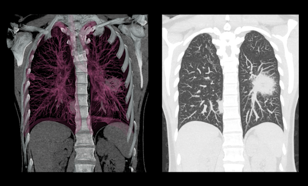
CT scans are preferred for staging tumors and are vital for patients with malignant pleural mesothelioma, Yendamuri explained. The 3-D images CT scans provide a far more detailed view and offer more than 90 percent detection sensitivity. However, appearances can be subjective and are highly operator dependent, he said. Furthermore, although less effective for detecting peritoneal mesothelioma, CT scans are still the most useful imaging study for diagnosing malignant pleural mesothelioma. Scans can suggest or rule out mesothelioma diagnosis completely, while also helping to estimate the extent or stage of mesothelioma. Information obtained from CT images will also help determine whether the tumor can or should be removed with surgery.
These are the common radiological findings in mesothelioma CT scans:
• Tumor extent along pleural surfaces and into the diaphragm, mediastinum, or chest wall
• Chest wall and diaphragmatic invasion
• Pleural thickening and effusion
• Nodular pleural thickening, pleural thickening greater than 1 cm, mediastinal pleural surface and concentric pleural thickening
• Calcified pleural plaques
Use of MRI in Mesothelioma
MRI exams complement CT scanning in some patients, providing better delineation of soft tissues and allowing imaging in the sagittal and coronal planes. MRI scans also enable better detection of mesothelioma metastasis, whereas CT scans are more likely to miss metastasis, Yendamuri said. MRI tests are also suitable for patients who cannot tolerate the contrast dyes used during CT scans. CT scans and MRI tests do not provide an unequivocal diagnosis of mesothelioma, so tissue biopsy is required for definitive diagnosis.
Common MRI findings include:
• Solitary foci of chest wall invasion, endothoracic fascial involvement, and diaphragmatic invasion.
• Iso-intense T1 signals of chest wall musculature
• Moderate increased signals on T2-weighted images or enhanced T1-weighted images when gadolinium is injected
Use of PET Scans for Mesothelioma
PET nuclear scans are helpful in determining the prognosis of patients with malignant pleural mesothelioma, as they are sensitive enough to detect small increases in metabolic activity, allowing the detection of extremely small collections of cancer cells in remote locations of the body. Resolution of PET scan images are relatively low, hence the use of dual-imaging combinations of PET and CT scans at most modern cancer centers. This imaging technology is less valuable in the diagnosis and staging of peritoneal mesothelioma, but can be helpful in specific instances.
Common PET findings include:
• Extent or stage of lymph node involvement, tumors or metastases
• Differentiation of pleural mesothelioma from benign pleural lesions
• Pleural inflammation
Diagnosis is Only a Small Portion Mesothelioma
With mesothelioma diagnosis being difficult to confirm, it is important to remember that diagnosis itself is only a small portion of the mesothelioma treatment timeline. Imaging tests are important for determining diagnosis and the stage of mesothelioma. Images can provide insights about which treatments might be most effective, Yendamuri said.
“Being aware of mesothelioma during the evaluation of a pleural effusion so that mesothelioma is diagnosed earlier should hopefully make available, more therapeutic treatment options,” Yendamuri urged.
Editor’s note: Alison Grimes is a health advocate for the Mesothelioma + Asbestos Awareness Center (www.maacenter.org). The center offers an online resource for patients
For more information on the disease visit mesothelioma.net
References:
1. William G. Bradley. (Sep 2008). Proceedings of the American Philosophical Society Vol. 152, No. 3 (Sep., 2008), pp. 349-361 American Philosophical Society. Retrieved from http://www.jstor.org/stable/40541591
2. Fazal Hussain, MD, MBBS Associate Professor, College of Medicine, Alfaisal University College of Medicine, Saudi Arabia Retrieved from (http://emedicine.medscape.com/article/359470-overview?pa=9V5hp7frfzbp7cv7rIfsNowbILvj1hXyMyxFeEcwjaUifaDkNvr5EeRl3d%2BZVybDLxY8ILgX%2Fhmc1eVgnkUOUON5lPYw%2FtQ7Z8WOOzpssmw%3D)
3. Anna C. Bibby1, 2, Selina Tsim3, 4, Nikolaos Kanellakis5, 6, Hannah Ball6, 7, Denis C. Talbot 7, Kevin G. Blyth3, 8, Nick A. Maskell1,2 and Ioannis Psallidas5,6 Number 4 in the Series “Pleural Diseases” Malignant pleural mesothelioma: an update on investigation, diagnosis and treatment. CrossMark Publishing. Retrieved from: (http://err.ersjournals.com/content/errev/25/142/472.full.pdf)
4. www.maacenter.org/mesothelioma/diagnosis/mesothelioma-imaging-tests/

 April 17, 2024


