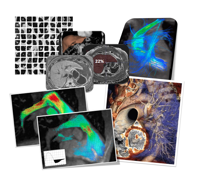
Collage depicts broad applications in machine learning or deep learning (DL) that can be applied to advanced medical imaging technologies. Size of the liver and its fat fraction — 22 percent — (top middle in collage) can be quantified automatically using an algorithm developed by Dr. Albert Hsiao and his team at the University of California San Diego. This and other information that might be mined by DL algorithms from CT and MR images could help personalize patients’ treatment. Collage provided by Albert Hsiao
Computed tomography (CT) and magnetic resonance imaging (MRI) scans are chock full of information that might be used to personalize treatment. Unfortunately, this information is not readily available. The latest type of artificial intelligence (AI) algorithms, however, might change that, according to Albert Hsiao, M.D., assistant professor of radiology at the University of California San Diego.
During a scientific session March 16 of this year’s annual meeting (#ACC19) of the American College of Cardiology, Hsiao said he plans to discuss how smart algorithms might extract this information from CT and MRI scans and how its availability could change patient management.
A key takeaway of his presentation, to be given as part of a scientific session titled “Man vs. Machine: Current and Future Applications of Machine Learning in Cardiovascular Imaging,” will be that the “explosive growth” of machine learning has raised “opportunities for tying (this information) directly to patient management,” he said. “This is in large part because of the amazing democratization of machine learning that this new area of ‘deep learning’ has enabled.”
Physicians might use this information to create “imaging phenotypes” of patients. These phenotypes, in turn, might be used to personalize the treatment of patients.
“Information like how much body fat you have, how fat is your liver, how much is your muscle mass — those are rich pieces of information that if quantified could really help us guide and make more specific patient recommendations,” said Hsiao, who seven years ago co-founded Arterys, a modern day provider of cloud-based AI software for medical imaging.
Imaging phenotypes would characterize the physical aspects of a patient, seen in medical images. For the time being the information remains embedded in these images, he said. But smart algorithms might extract and quantify it. Doing so could give clinicians insights into exactly what their patients need, allowing an unprecedented degree of personalization.
DL Growth To Be Cited at #ACC19
Deep learning (DL), also called machine learning or unsupervised learning, allows algorithms to discover rules for distinguishing certain features of images. In the early days of AI, DL allowed some algorithms to distinguish pictures of cats from those of dogs. Rather than coding the rules for distinguishing these pictures, the algorithms “learned” them.
Today’s DL algorithms might similarly learn how to pick out and extract phenotypic information from CT and MR images. They would do so by leveraging convolutional neural networks (CNNS), which are typically used to analyze images.
Deep learning — and CNNs — might be applied in any medical imaging modalities, including echocardiography and X-ray angiography. But the three-dimensional nature of CT and MRI make these two modalities attractive, especially when it comes to AI efforts aimed at quantitation, Hsiao said, for example, determining the fat fraction of a liver.
Over the past eight years, Hsiao has co-authored articles primarily on smart algorithms in cardiac MRI. But there is also “quite a bit of open terrain” regarding CT, Hsiao said: “There's some interesting work for coronary CT and estimating CT-FFR (fractional flow reserve) with machine learning.”
How To Generalize Algorithms
The challenge facing AI developers is to validate these algorithms across multiple patient populations. Algorithms typically are developed and tested in a single population type. This may be exemplified by a cohort of 200 to 300 patients. Achieving “generalizability” may be especially important when trying to dig information out of CT and MR images — information needed to classify patients according to “imaging phenotypes” — because different types of patients may express different types of imaging phenotypes.
The challenge is not, however, as difficult as might be imagined, Hsiao said: “We just basically need the data from these other demographics.”
The data are required, he said, not only to refine the algorithms but to prove that they work on different types of patients. This proof is needed if the algorithms are to have wide clinical utility — and, consequently, to establish the value of unused data now buried in CT and MR images.
Greg Freiherr is a contributing editor to Imaging Technology News (ITN). Over the past three decades, Freiherr has served as business and technology editor for publications in medical imaging, as well as consulted for vendors, professional organizations, academia, and financial institutions.
Related content:
Applications for Artificial Intelligence in Cardiovascular Imaging
Machine Learning Approaches in Cardiovascular Imaging
Technology Report: Artificial Intelligence (Video report published January 2019)
PODCAST: Shortcomings of CTA in Cardiology
PODCAST: How Technology Is Changing Cardiology
ACC.19 Future Hub Hosts “Shark Tank” of Emerging Technologies In Cardiology


 April 17, 2024