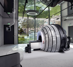
The InSightec ExAblate focused ultrasound system developed to treat essential tremor. Studies are underway to see if FUS can be used to treat other brain conditions and brain cancer.
Focused ultrasound (FUS), also called high-intensity focused ultrasound (HIFU), has gained a lot of interest in recent years as noninvasive cancer therapy that does not require radiation, chemotherapy, or surgery, resulting is less collateral damage to the patient. FUS is performed as an out-patient procedure, so it can eliminate the usual two weeks of recovery needed from most types of surgery.
In the U.S., the U.S. Food and Drug Administration (FDA) has cleared FUS to treat bone metastases, uterine fibroids, prostate cancer, benign prostatic hyperplasia and essential tremor. Only bone metastases currently has reimbursement. Outside the U.S., there are additional regulatory approvals for breast cancer, kidney cancer, liver cancer, pancreatic cancer, soft tissue tumors, Parkinson’s disease, thyroid nodules, back pain, osteoid osteoma, breast fibroadenomas, uterine adenomyosis and neuropathic pain.[1]
Currently, use of FUS is generally considered only if it will have a greater impact on the patient than surgical outcomes, said Pejman Ghanouni, M.D., Ph.D., assistant professor of radiology at the Stanford University Medical Center. He is an expert in FUS and involved in research for the use of FUS in the treatment, including essential tremors, soft tissue tumors and localized low-intermediate risk prostate cancer. He spoke on the topic at sessions at the Radiological Society of North American (RSNA) 2016 annual meeting.
As more providers begin offering FUS treatment programs, Ghanouni said it is important to have a radiologist champion for the project. Patient recruitment from referrals also needs to be considered, so it is important to build relationships with referring physicians. For example, at Stanford he said they have a fibroid center, and one in 10 of the patients with uterine fibroids will qualify for FUS treatment. He said this is how his center primed the pump for initial referrals. He also now works with Stanford’s prostate cancer center, radiation therapy and desmoid tumor clinic. He said one of the keys to finding patients has been defining unmet needs and how FUS can serve as a niche treatment option.
The Basics of High-Intensity Focused Ultrasound
According to the Focused Ultrasound Foundation, the principle of FUS is analogous to using a magnifying glass to focus beams of sunlight on a single point to burn a hole in a leaf. With focused ultrasound, an acoustic lens is used to concentrate multiple intersecting beams of ultrasound on a target deep in the body with extreme precision. Depending on the design of the lens and the ultrasound parameters, the target can be as small as 1 x 1.5 mm or as large as 10 x 16 mm in diameter. Where each of the individual beams passes through the tissue, there is no effect. But, at the focal point where these multiple beams converge, the focused ultrasound energy results in tissue ablation.[2]
FUS uses thermal ablation to denature proteins and cause cell death. The thermal dose required to produce irreversible damage and coagulative necrosis depends on the cell type, temperature and duration of exposure. This ranges from one second at 130°F, to 240 seconds at 107°F. FUS also uses mechanical tissue destruction to disrupt cells through cavitation, in which bubbles of gas oscillate in an ultrasonic field and collapse which can generate enough force to allow for the targeted destruction of tissue.[3]
FUS works in conjunction with magnetic resonance imaging (MRI), which is used to identify and target tissue to be treated, for real-time image guidance and control during treatment, and to confirm the effectiveness of the therapy. MRI is used to create a treatment plan similar to radiation therapy. A post-procedure MRI also is performed to confirm the effectiveness to the treatment. For this reason, hospitals interested in creating FU programs need staff who are comfortable working in the MRI suite. The close integration with MRI often has also lead to the therapy being referred to as magnetic resonance-guided focused ultrasound (MRgFUS).
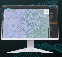
The treatment plan for FUS used the anatomical MR imaging with an overlay of the zones of treatment. These are shown as strips, each representing a pass of the FUS beam. These strips are usually stacked on top of each other and the length of each varies to match the contours of the tumor target.
Operators need to watch the real-time MR imaging for the formation of bubbles in the tissue being ablated, which can deflect the FUS beams.
FUS of Uterine Fibroids and Avoiding Complications
MRI anatomical assessment is used at the start of patient evaluation to determine if the target is reachable by FUS, said Young-Sun Kim, M.D., assistant professor, Department of Radiology and Center for Imaging Science, Samsung Medical Center, Seoul, South Korea. He is an expert in FUS and spoke RSNA 2016 sessions. In the case of uterine fibroids, he showed an example where the treatment window to access the fibroid might be blocked by cysts in the path of the ultrasound beam. He said thick subcutaneous fat is another ultrasound beam attenuation factor. Edema caused by fluid back up in a fibroid may also lead to poor outcomes with FUS. He also suggests avoiding resistant fibroids.
Additionally, Kim radiologists need to assess MR imaging along the planned trajectory of the beams to look for foreign bodies, which often present as surgical procedure left overs like staples. He said these can superheat during the procedure and injure the patient.
MRI offers real-time thermography during the FUS treatment, to ensure health tissue are not damaged. Kim said complications from FUS can include skin burns, fat burns, sciatic nerve injury and bowl injury. Like radiation therapy, Kim said the focused ultrasound beams also can cause damage to critical structures, such as nerves and the spine. This is why he suggests using conscience sedation with IV fentanyl so patients can offer feedback during the procedure.
Critical structures like the bowel and spine can often be avoided by manipulating the bladder of bowel. Kim said this can be done by filling or emptying the bladder, or filling the bowel with ultrasound gel to move the bowel out of the way, or to to eliminate bowel loops.
Treatment of Bone Tumors, Metastasis
As tumors grow inside bones, they slowly destroy and break apart the bone as the tumor expands. With osteoid osteoma, benign bone tumors, Napoli said HIFU can both reduce the pain and stop the tumor growth. However, with bone metastasis it is best to limit treatment to the palliation of pain, said Alessandro Napoli, M.D., Ph.D., Department of Radiological and Oncological Sciences, Sapiens University, Rome, who spoke on FUS at RSNA 2016.
“It is totally noninvasive, there is no radiation and we have low complication rates compared to other therapies. This is because it is totally image guided,“ Napoli explained. “The quality of life is improved greatly with the reduction of pain.”
During the procedure, he uses an epidural nerve block or deep sedation because of the pain involved in the procedure. His center also uses gel pads to act as a coupler between the FUS and the body.
He uses perfusion MRI with contrast both before and after procedures so he can see reductions in tumor vascularity.
Napoli said you can direct FUS beams through nerves if the beam is wide to reduce damage. The bone also absorbs a lot of the FUS so the treatment time will depend on the density of the bone and amount of bone that needs to be penetrated.
FUS does not hamper the bone growth, which Napoli said has been proven in several studies. Imaging from these studies has shown new bone growth months or years later with restored bone.
Read the article "Researchers Investigate Using Focused Ultrasound To Take Away Cancer Pain."
FUS of the Brain
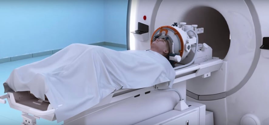
A growing area of HIFU is in treatment of essential tremor by applying FUS for thalamic ablations. In this application, a special spherical FUS transducer system has been developed that fits over the patient’s head. Ghanouni showed before and after videos of patients he treated who could not hold a glass of water without spilling it, or use a spoon. After treatment they were able to drink from the glass or use the spoon normally.
Brain FUS uses an array of about 1,000 elements designed to overcome attenuation issues presented by the skull and to diffuse the heating over a large area of the brain to prevent damaging healthly tissue.
“Bone is very efficient at absorbing ultrasound energy,” Ghanouni said, explaining why so many ultrasound elements are needed to treat the brain and avoid overheating. Additionally, a silicon pad surrounding the head that circulates chilled water is used to transfer the ultrasound beams into the skull and to cool the skin and head surface. Peri-procedural MR thermography is used to monitor temperatures and ensure areas outside of the target are not being heated. The patient’s head also is shaved to permit the transference of ultrasound waves.
In the case of the brain, he said computed tomography (CT) is used for registration in the treatment planning system. Planning includes avoiding any calcifications inside the brain, because they will heat up during treatment, Ghanouni said.
The procedure is performed with conscience sedation so the patient can offer intra-operative physiologic feedback. When the operators isolate the target area, they first perform sub-lethal sonifications to confirm their positioning before delivering lethal sonifications.
These brain ablations can cause edema, but Ghanouni said this usually resolves over time, and as they reduce in size, there usually is dramatic improvement in the patients. “The first week is not representative of what the final result will be,” he explained, adding there can be some bad side effects, but these often resolve or change after about three months. “The final results is often not seen until after the three month recovery period, so patents need to be told this upfront. What they have after three months is usually what they will keep.”
He said the FDA recently approved FUS for essential tremor and there is an expectation that CMS may begin reimbursing for the FUS sometime in 2017.
Ghanouni said a next step will be FUS treatment of Parkinson’s disease, which is already an approved indication overseas.
Another use of FUS in the brain may be to improve targeted drug delivery to specific tumors. Ghanouni said FUS can help break down the blood-brain barrier with the use of microbubbles. Early studies show this breakdown of the barrier only lasts a couple hours before it disappears.
HIFU to Treat Prostate Cancer
Napoli said HIFU can be used as a primary treatment for prostate cancer, or as a salvage treatment when there is recurrence after surgery or radiation therapy. He said it is important to target the individual lesions, not to use FUS for whole gland treatment, which can lead to urinary incontinence and erectile dysfunction.
He said a special FUS transrectal transducer has been created so it can be placed against the rectal wall, next to the prostate. This helps focus on the prostate without worry of causing collateral damage to surrounding critical structures.
“The primary argument against targeted treatment for the prostate is that the disease is multifocal,” Napoli explained. He said this often requires the excision of the whole gland. “However, in most cases, a single index or dominant lesion, drives prostate cancer risk. The optimal candidates for targeted ablation need to be clearly identified.”
Unlike surgery or brachytherapy, HIFU is significantly less invasive, which may be much more appealing to patients. “It does not require incisions or punctures, it is bloodless, can be carried out on an outpatient basis, and it is repeatable,” Napoli said.
Future Directions for HIFU
The use of FUS is still relatively new and there are many pre-clinical and early stage pilot clinical programs investigating its expanded use for other conditions. Expansion in oncology may include several new cancers in the coming years. Pilot trials have started for pediatric neuroblastoma, melanoma, brain, head and neck, lung, ovarian and cervical cancers.
In neurology there are pilot clinical studies for brain tumors, depression and obsessive-compulsive disorder (OCD). Preclinical studies are underway for Alzheimer’s disease, epilepsy, multiple sclerosis, stroke, traumatic brain injury and trigeminal neuralgia.
Read the article "Pre-Clinical Research Validates Potential for Focused Ultrasound in Alzheimer's."
In cardiovascular medicine, there are pilot trials testing FUS to treat hypertension. There also are numerous preclinical studies looking at the use of FUS to treat arteriovenous malformations, atherosclerosis, atrial fibrillation, deep vein thrombosis, heart block, peripheral artery disease (PAD), septal perforation and heart failure.
References:
1. Focused Ultrasound Foundation. “Diseases and Conditions.” www.fusfoundation.org/diseases-and-conditions/overview. Accessed Feb. 2, 2017.
2. Focused Ultrasound Foundation. “Technology Overview.” www.fusfoundation.org/the-technology/overview. Accessed Feb. 2, 2017.
3. Focused Ultrasound Foundation. “Mechanisms of Action” www.fusfoundation.org/the-technology/mechanisms-of-action. Accessed Feb. 2, 2017.
4. Alessandro Napoli, Pejman Ghanouni, Young-Sun Kim. “Emerging Technology: High Intensity Focused Ultrasound - Opportunities and Challenges.” RSNA 2016 CME session, RC617. Presented Thursday, Dec. 1, 2016.


 April 18, 2024