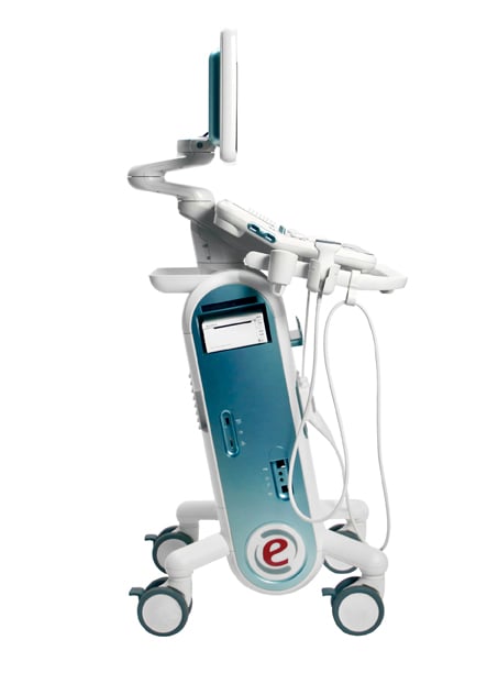
Esaote's MyLab Six.
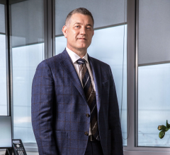
The future of ultrasound looks auspicious, according to Jonathan M. Rubin, M.D., Ph.D., director of the division of ultrasound in the department of radiology at the University of Michigan in Ann Arbor, who presented on “The Future of Ultrasound” at the 2014 Radiological Society of North America (RSNA) conference in Chicago this past December. His lecture highlighted emerging techniques for volume blood flow and shear wave imaging.
Rubin explained that the use of 3-D and 4-D ultrasound using an angle-independent technique for flow estimation is now possible, and quite simple to measure. “The technique is based on Gauss’s Theorem. This relation states that volume flow is equal to the total integrated flux over any surface cutting across a conduit flow. The method is angle independent, flow profile independent and vessel geometry independent,” he stated.
Using 2-D ultrasound arrays, these measurements could be performed in real time, said Rubin. “A good approximation of its potential utility corresponds to the parameters such as resistive indices and pulsatility indices that are now being applied,” Rubin explained, noting that volume flow could replace these. The measurements could affect analyses such as transplant evaluations, cardiac output measurements, fetal evaluation through umbilical cord blood flow measurements, carotid artery flow and cerebral perfusion. “Direct perfusion estimates defined by flow per unit mass would become standard by estimating organ weight with 3-D imaging and direct measurements of blood flow.”
Elasticity imaging has become another new approach to ultrasound, with the general execution of elasticity imaging being strain and shear wave speed (SWS) imaging. Currently, both are augmented by shear viscosity imaging, nonlinear strain and nonlinear shear wave imaging.
The topic generating the most conversation is assessment of liver fibrosis/cirrhosis, since the current standard of care for evaluating the liver is through biopsy. “Biopsies are also highly localized and represent a very poor sample of the underlying disease. SWS imaging is a more global measure. For these two reasons, SWS will likely replace liver biopsies for fibrosis/cirrhosis assessment,” said Rubin.
Other ultrasound applications could assist with breast cancer differentiation, thyroid nodule characterization, cardiac function and conduction analysis, deep vein thrombosis aging and Crohn’s disease stricture evaluation.
Ultrasound System Improvements
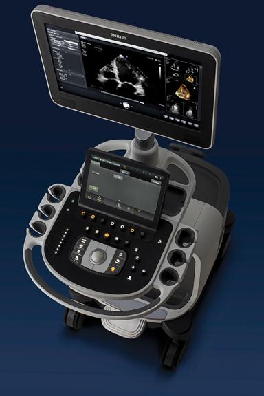
At RSNA, Philips introduced an ultrasound consortium to drive the creation of mobile ultrasound solutions. This includes an app-based ultrasound system, which Philips demonstrated at its booth. It uses a transducer capable of connecting to a mobile device and a downloadable app to turn a consumer-grade smartphone of tablet device into an ultrasound machine. The product was developed for market testing and research and is not currently for sale.
Philips also showcased several new ultrasound systems, including Epiq 5 and 7, and the Affiniti. The Epiq 5 and 7 utilize PureWave technology, allowing for improved penetration in difficult patients with a single transducer. New elastography imaging capabilities assist in acquiring additional data for liver disease diagnosis using liver stiffness measurements rather than liver biopsies. This technique is performed for assessments of diffuse liver diseases and neoplastic lesions of the liver.
Ergonomics, workflow and mobility improvements are touched upon as companies find more ways to improve their ultrasound machines. Philips data states that more than 80 percent of ultrasound users experience work-related pain, with more than 20 percent of these users suffering a career-ending injury. The ultrasound systems can be used sitting or standing, and can be transported on carpet and tile. Its touch interface provides 40 percent less reach and 15 percent fewer steps to complete an exam. The xMatrix on the Epiq 7 contains seven modes in a single transducer: 2-D, 3-D/4-D, Live xPlane, Live MPR, Doppler, color Doppler and color power angio (CPA).
Affiniti was produced to enable global hospitals and healthcare systems to overcome the demands of increasing patient volumes and cost pressures. It debuted at the European Society of Cardiology Congress 2014 (ESC) in Barcelona, Spain, and was built on the same platform as the Epiq systems. PureWave transducer technology is incorporated in the system, countering the challenges of imaging obese patients and those with liver disease. The PureWave transducer works with the Affiniti ultrasound system to accommodate for the altered speed of high-frequency sound waves through adipose layers versus other tissue, according to Philips. This helps the ultrasound system become aware of increased adipose content. It then applies aberration correction of algorithms, which results in a high-quality image throughout the entire beam length. Users reported exam times were decreased 30-50 percent when using Affiniti.
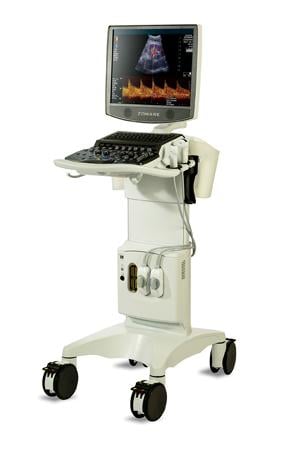
Also at RSNA, Zonare Medical Systems announced upgrades for its ZS3 ultrasound platform in image quality, transducer technology, workflow and clinical enhancements. The technology has the ability to utilize all information in the returning echo data set with sufficient coverage of the imaging field of view occurring in fewer transmit/receive cycles. Unlike conventional beamformer line-by-line imaging, Zonare uses large zone acquisitions. Users can view examinations in grayscale and color Doppler, and retention of channel domain data make patient-specific imaging possible. Retrospective imaging captures raw data with the ability to process it multiple times and ways. Nineteen transducers are available for ultrasound use, one of which contains a hand-held section for operators’ fingers, making it less cumbersome and more comfortable to conduct scans.
Esaote showcased its Virtual Navigator fusion imaging at RSNA 2014, which recently received U.S. Food and Drug Administration (FDA) clearance. Virtual Navigator fuses real-time ultrasound images with magnetic resonance (MR), computed tomography (CT), positron emission tomography (PET) and 3-D ultrasound. Urologic, musculoskeletal, peripheral vascular and transcranial radiologic exams can be performed, and it also is utilized for liver and other abdominal interventions.
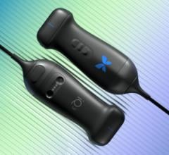
MyLab Six and MyLab Gamma, Esaote’s newest ultrasound systems, were developed to meet demands of the sonographer’s working environment. MyLab Six’s ergonomics are apparent in the slanted body of the machine so users do not hit their knees or feet on the machine. The cart-based system has a keyboard that rises up or down depending on user preference. Images and data can be seen on a 19-inch LCD monitor that was designed to reduce eyestrain. Settings on the machine can be customized and organized based on clinical preferences. The noiseless system allows sonographers the ability to focus on the exam and diagnosis with minimal distraction. The “green” ultrasound system uses less power, reducing costs to run the machine. Another ergonomic feature is the iQProbe appleprobe, shaped to keep the hand and wrist in a natural grip, preventing tension. The probe delivers an ultrasound beam and receives a backscattered echo. The technology is important in obtaining a high signal-to-noise ratio to optimize information regarding spatial, contrast and temporal resolution.
MyLab Gamma is a portable laptop-sized ultrasound system that can be transported between departments within a hospital or clinic. The system is battery operated and light enough to transport on a user’s back. The monitor can be rotated through +90/-90 degrees, which allows examinations to be shared with patients and colleagues. The eTouch screen-based interface guides operators through setup and image capture, which enables a faster workflow. The iQProbe appleprobe is also available in this system.
What’s Next
Ultrasound is used by a myriad of imaging technologists because it is considered practical, safe, fast and easy to utilize. With new advancements in ultrasound technology, many more patients and users can benefit from reduced costs, shorter exam times and eliminating the need for additional exams using other modalities.
“These mainly passive attributes will be and are being augmented by new quantitative methods that are nearly unique to ultrasound, giving ultrasound a new life and an increased relevance in the medical imaging armamentarium,” Rubin concluded.
Access the most current version of the ITN Ultrasound Systems Comparison Chart (www.itnonline.com/content/ultrasound-systems). This will require a login, but it is free and only takes a minute to complete the form.
Read the 2016 article “Top Trends in Cardiovascular Ultrasound.”
Read the 2016 article “Ultrasound Enhances Quality as Technology Advances.”
Watch the VIDEO: Trends in Ultrasound at RSNA 2016

 April 08, 2024