itnTV

VIDEO: One on One with Amy K. Patel, MD, American Association for Women in Radiology Immediate Past President

Breast Imaging | April 15, 2024

Don't miss ITN's latest "One on One" video interview with AAWR Past President and American College of Radiology (ACR) RAN and RADPAC Chair, Amy K. Patel, MD, discussing advocacy initiatives and innovations in artificial intelligence (AI) for breast imaging

Dr. Patel is a breast imaging trailblazer and radiology advocacy leader. In this video,  learn how radiologists can support key initiatives, ways AI is improving patient care, and more.

Related content:

Leaders from RadEqual and the AAWR Sign MOU, Solidifying Commitment to Advance Opportunities in Radiology

Technology Report: Artificial Intelligence in Radiology 2021

VIDEO: Integrating Artificial Intelligence Into Radiologists Workflow

Recent Video

Radiation Therapy | February 02, 2022

Magdalena Bazalova-Carter, Ph.D., assistant professor, University of Victoria University, discusses the current state of ultra-high dose FLASH radiation therapy at the 2021 American Society of Radiation Oncology (ASTRO) annual meeting. Flash therapy is said to be a key technology to keep an eye on in the next few years. If it proves viable in human patients, it promises to greatly shorten treatment times, and reduce fractions to between 1-3 sessions.

The idea is that a super-high dose of radiation is delivered in one large, very fast dose. It appears that despite the high dose of radiation, there is a tissue sparing biology mechanism that is not yet fully understood, where health tissue is preserved and there is less collateral damage than the standard series of lower dose fractions over days or weeks.  

Flash therapy is being tested in electron beam therapy systems to treat superficial cancers, which are much easier to adopt to flash than deeper tissue tumors. Proton may be able to produce the higher energies needed for deeper tumor treatments, but current photon beam systems are limited because to deliver the high doses needed may cause enough heat to melt the X-ray beam source.

7 Trends in Radiation Therapy at ASTRO 2021

Photo Gallery of Technologies at ASTRO 2021

Radiation Oncology Research Featured at ASTRO 2021

Find more radiation oncology technology news

Radiation Oncology | February 02, 2022

Brian S. Bingham, M.D., chief resident in radiation oncology at the Vanderbilt University Medical Center, explains the practice-level and national cost burden of treatment-related prior authorization for academic radiation oncology practices. This was a highlighted study presented at the 2021 American Society of Radiation Oncology (ASTRO) annual meeting.

Photo Gallery of Technologies at ASTRO 2021

Radiation Oncology Research Featured at ASTRO 2021

Find more radiation oncology technology news


Radiation Therapy | February 02, 2022

Nima Nabavizadeh, M.D., radiation oncologist and associate professor of radiation medicine, Oregon Health and Science University, explains the use of external-beam radiation therapy as a bridge to transplant in hepatocellular carcinoma cancer patients.

He presented a utilization analysis of the United Network for Organ Sharing (UNOS) database at the 2021 American Society of Radiation Oncology (ASTRO) annual meeting.

7 Trends in Radiation Therapy at ASTRO 2021

Photo Gallery of Technologies at ASTRO 2021

Radiation Oncology Research Featured at ASTRO 2021

Find more radiation oncology technology news

Radiation Oncology | February 02, 2022

Bridget F. Koontz, M.D., chief medical officer at GenesisCare USA explains how to manage the complex needs of pelvic radiotherapy survivors. She offers an. overview of published evidence about the various toxicity types and approaches for managing them.

Photo Gallery of Technologies at ASTRO 2021

Radiation Oncology Research Featured at ASTRO 2021

Find more radiation oncology technology news

7 Trends in Radiation Therapy at ASTRO 2021


Radiation Oncology | February 02, 2022

Douglas E. Holt, M.D., a radiation oncologist at Eastern Idaho Regional Medical Center, explains the use of 3-D virtual reality volumetric imaging review to help improve cancer patients’ understanding of their disease and treatment. Pictures are worth a thousand words, and moving pictures inside a patient's body even more. Holt said using virtual reality to go through the patient's anatomy in 3D and to show them what is wrong and how it will be treated offers a new level of understanding that is not possible using a discussion or a couple still images from their medical imaging.

Holt presented this study as a late-breaker at the 2021 American Society of Radiation Oncology (ASTRO) annual meeting.

Find more ASTRO videos and news

7 Trends in Radiation Therapy at ASTRO 2021

Photo Gallery of Technologies at ASTRO 2021

Radiation Oncology Research Featured at ASTRO 2021

Find more radiation oncology technology news

Quality Assurance (QA) | January 31, 2022

Sun Nuclear highlighted two resources for CT Quality Assurance at RSNA 2021. ITN visited their booth to learn more about the Mercury 4.0 Phantom and the Multi-Energy CT Phantom from Thomas Webb, Global Product Marketing Manager

Find more RSNA news and videos

Digital Radiography (DR) | January 31, 2022

Agfa recently announced the launch of the new Valory digital radiography room, which was designed for health care facilities requiring a highly productive radiography solution. 

ITN stopped by the Agfa booth at RSNA 2021 to learn more about this new system from Georges Espada, Head of Digital Radiography, and Karol Wesolowski, Global Category Leader.

Coronavirus (COVID-19) | January 31, 2022

Marc Succi, M.D., an emergency radiologist at MGH and executive director of the MESH Incubator, an in-house innovation and entrepreneurship center, and  Ottavia Zattra, a fourth-year medical student at Harvard Medical School, explain a study they authored showing there might be higher cancer rates due to lower numbers of CT scans during COVID-19. They presented this study as a late-breaker at the Radiological Society of North America (RSNA) 2021 annual meeting.

COVID caused many people to delay seeing their doctors. Their study found a corresponding 82% drop in CT imaging for cancer screening. CT is also used for initial cancer workups, to monitor active cancer and post procedure surveillance, which all also showed decline since the start of the pandemic.

Read more about the study COVID-19 Fallout May Lead to More Cancer Deaths

Computed Tomography (CT) | January 28, 2022

Charlie Hamm, M.D., a radiology resident at the Charité University Hospital of Berlin, Germany, presented a late-breaking study at the Radiological Society of North America (RSNA) 2021 annual meeting on the use of computed tomography (CT) scans to investigate dinosaur bones non-destructively. In the process of examining a tyrannosaurus rex jaw bone that is more than 66 million years old, a bone tumor was found and clearly shown on the CT scans.

This feasibility study to determine if CT can be used to aid paleontology was done in collaboration with the Museum für Naturkunde Berlin. Dual-energy computed tomography (DECT) was used to provide information about tissue composition and disease processes not possible with single-energy CT.

Read more in the article CT Uncovers Bone Disease in Tyrannosaurus Rex Jaw

Find more RSNA news and video


Quality Assurance (QA) | January 28, 2022

Mahadevappa Mahesh, Ph.D., chief physicist, Johns Hopkins Hospital, and professor of radiology and radiological science, explains the basics involved in quality assurance (QA) of radiology imaging systems. He spoke to ITN at the Radiological Society of North America (RSNA) 2021 annual meeting.

He explains the role of the medical physicist in keeping X-ray imaging systems such as CT, angiography and mammography calibrated and checking the device output of radiation. This is performed by imaging phantoms that mimic a simplified representation of the human body. 

Artificial intelligence (AI) use is growing in imaging and medical physics and QA of these systems might also become a duty of the medical physicist in some AI imaging applications.  

Find more content on QA systems

Find more RSNA news and video

Artificial Intelligence | January 27, 2022

Emanuel Kanal, M.D., FACR, FISMRM, AANG, director of the department of emergency radiology and teleradiology, director of MRI services, and professor of radiology and neuroradiology at the University of Pittsburgh, explains artificial intelligence (AI) is the biggest over all trend in radiology at  the Radiological Society of North America (RSNA) 2021 annual meeting.

VIDEO: Artificial Intelligence Trends in Medical Imaging — Interview with Sanjay Parekh, Ph.D.

VIDEO: Examples of Artificial Intelligence Pulmonary Embolism Response Team Apps

Technology Report: Artificial Intelligence in Radiology 2021

Find more AI news

Find more RSNA news and video

Enterprise Imaging | January 27, 2022

Rik Primo, principle at Primo Medical Imaging Informatics Consultants and former health IT developer with Siemens, Philips and Agfa, explains the difference cloud-native versus cloud-enabled PACS and radiology enterprise imaging systems. He spoke with ITN during RSNA 2021.the Radiological Society of North America (RSNA) 2021 annual meeting.

Find more RSNA news and video

Related content on enterprise imaging


Computed Tomography (CT) | January 27, 2022

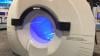
Cynthia McCollough, Ph.D., director of Mayo Clinic's CT Clinical Innovation Center,  explains how photon-counting computed tomography (CT) detectors work and why it is a better technology over conventional CT systems. She helped Siemens develop the Naeotom Alpha, the first photo-counting CT system to be approved by the FDA in the fall of 2021. She spoke to ITN at the Radiological Society of North America (RSNA) 2021 annual meeting.

Read more about the first commercial photon-counting scanner 

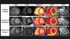
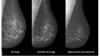
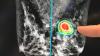
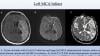
The device uses the emerging CT technology of photon-counting detectors, which can measure each individual X-ray photon that passes through a patient's body, as opposed to current systems which use detectors that measure the total energy contained in many X-rays at once. By "counting" each individual X-ray photon, more detailed information about the patient can be obtained and used to create images with less information that is not useful, such as image noise. 

Current CT technology uses a two-step conversion process to convert X-ray photons into visible light using a scintillator layer in the detector. Then, photo diode light sensors turn the visible light into a digital signal. Due to this intermediate step, important information about the energy of the X-rays is lost and no longer available to aid in diagnosis. Also, contrast is reduced and images are not as clear.

Photon-counting detectors use a single step of direct conversion of X-rays into electrical current, and skips the step of converting X-rays into visible light. This allows the energy thresholds of each pulse to be collected and binned based on different kilovolt (kV) energy levels. This creates data to improve contrast and enable dual-energy, spectral imaging. The direct conversion also helps improve image quality without information loss. This improves image sharpness and contrast.

Photon-counting detectors have already been used for several years in high-energy physics and nuclear imaging. However, these previously generation photon-counting detectors could not be used with a clinical CT scanner because they could not keep up with the high higher rate of photons reaching the detector. The detector on the Naeotom Alpha was designed for this increased speed.

Related Photon-counting CT Content:

Mayo Clinic Begins Use of Third-Generation Photon-counting CT Clinical Research Detector

VIDEO: New Advances in CT Imaging Technology — Interview with Cynthia McCollough, Ph.D.

VIDEO: Photon Counting Detectors Will be the Next Major Advance in Computed Tomography — Interview with Todd Villines, M.D.

Key Trends in Cardiac CT at SCCT 2020

GE Healthcare Pioneers Photon Counting CT with Prismatic Sensors Acquisition

Top Trend Takeaways in Radiology From RSNA 2020

NeuroLogica Joins Forces with Massachusetts General Hospital to Pilot Photon Counting CT at the Patient’s Point-Of-Care Using OmniTom Elite CT

VIDEO: Advances in Cardiac CT Imaging — Interview with David Bluemke, M.D.

Computed Tomography (CT) | January 27, 2022

Cynthia McCollough, Ph.D., director of Mayo Clinic's Computed Tomography (CT) Clinical Innovation Center, explains how CT dose tracking software works and offers advice to centers that record this patient level and device information. She spoke to ITN at the Radiological Society of North America (RSNA) 2021 annual meeting.

Dose tracking software allows hospitals and imaging centers to track what levels of radiation they are using by exam type protocol. It can show technologists who are using higher than required doses that may need additional ALARA training. The radiation dose tracking systems also can help track the amount of radiation a patient has received over time.

Related Radiation Dose Tracking Systems:

Disputed EHR Dose Levels Could Keep Patients From Necessary Imaging Exams

Medical Imaging Radiation Exposure in U.S. Dropped Over Past Decade

VIDEO: Radiation From Medical Imaging in U.S. Dropped Over Past Decade

The Basics of Radiation Dose Monitoring in Medical Imaging

VIDEO: Radiation Dose Monitoring in Medical Imaging — Interview with Mahadevappa Mahesh, Ph.D.
 

----


Magnetic Resonance Imaging (MRI) | January 24, 2022

With the recent launch of the Magnifico Open, Italian company Esaote has entered the open MRI whole body space. ITN had a conversation with Franco Fontana, CEO of Esaote, at RSNA21.

Magnifico Open, which adds to the range of Esaote products unveiled in 2021, is an open magnetic resonance system with the latest technology. The wide choice of receiver coils and state-of-the-art MRI technology offer the user excellent image quality, while the permanent magnet makes it easy to use and lowers operating costs. The open magnet and the easy-to-access patient table also facilitate, speed up and make patient positioning more comfortable, ideal for both the claustrophobic and for children.

View more RSNA21 content here

Esaote North America Receives FDA Approval of the Magnifico Open MRI System

Magnetic Resonance Imaging (MRI) | January 18, 2022

Orlando Simonetti, Ph.D., professor, cardiovascular medicine, worked with Siemens to help develop a new, lower-field magnetic resonance imaging (MRI) system, the Magnetom Free.Max. It can scan patients that previously may have been contraindicated because of implantable medical devices. One of the first systems installed in the U.S. is at The Ohio State University Wexner Medical Center. It has a much lower magnetic field and a larger patient opening, removing barriers to MRI imaging for many patients.

Simonetti and his colleagues developed new techniques to boost the signal-to-noise ratio in MRI machines, which allowed the creation of a machine with a lower magnetic field strength that still enables high quality images.

The system gained FDA clearance in July 2021 and was featured by Siemens at the Radiological Society of North America (RSNA) 2021 meeting.

The interview and footage was provided by The Ohio University State University Wexner Medical Center.

Read more in the articles New FDA-approved MRI Expands Access to Life-saving Imaging and Ohio State Researchers Help Design New MRI, Expanding Access to Life-saving Imaging.

Related MRI Content:

Siemens Healthineers Announces First U.S. Installation of Magnetom Free.Max 80 cm MR Scanner

FDA Clears Siemens Healthineers Magnetom Free.Max 80 cm MR Scanner

Ohio State Researchers Help Design New MRI, Expanding Access to Life-saving Imaging


Artificial Intelligence | January 13, 2022

Here are two examples of artificial intelligence (AI) driven pulmonary embolism (PE) response team apps featured by vendors Aidoc and Viz.AI at the 2021 Radiological Society of North America (RSNA) 2021 meeting.

The AI scans computed tomography (CT) image datasets as they came off the imaging system and looked for evidence of PE. If detected by the algorithm, it immediately sends an alert to the stroke care team members via smartphone messaging. This is done before the images are even loaded into the PACS. The radiologist on the team can use a link on the app to open the CT dataset and has basic tools for scrolling, windowing and leveling to determine if there is a PE and the severity. The team can then use the app to send messages, access patient information, imaging and reports. This enabled them all to be on the same page and can communicate quickly via mobile devices, rather than being required to use dedicated workstations in the hospital. 

Both vendors showed similar apps for stroke at RSNA 2019. That idea for rapid alerts, diagnosis and communications for acute care teams has now expanded to PE and also for aortic dissection and abdominal aortic aneurysms (AAA). AI.Viz and Aidoc are looking at expanding this type of technology for other acute care team rolls, including heart failure response. 

Read more about this technology in the article AI Can Facilitate Automated Activation of Pulmonary Embolism Response Teams.

Find more AI news

Find more RSNA news and video

Enterprise Imaging | January 13, 2022

Steve Holloway, company director at Signify Research, explains the trends he has seen over the past couple years in enterprise imaging. He spoke to ITN at the 2021 Radiological Society of North America meeting.

Holloway shared how medical imaging systems are expanding to include all departments in healthcare system enterprises that generate data, images and waveforms, so these items can be stored in a central location, rather than disparate silos or in separate systems requiring multiple logins or specific workstations. Most of these systems are are web enabled or web based, allowing users to work from anywhere as long as they have an internet connection. Most enterprise imaging systems also use a web-based vendor neutral archive, allowing DICOM and non-DICOM images to be stored there. All of these features allow easier and faster access to patient information and images.

He said these systems are becoming more inclusive of ologies outside of radiology and cardiology. Most notably is digital pathology, which was featured by many enterprise imaging vendors at RSNA 2021. 

Enterprise imaging systems are also accepting point-of-care ultrasound (POCUS), which has exploded in use over the past two years with COVID, Holloway said.

Find more RSNA news and video

VIDEO: Trends in Radiology IT seen at RSNA 2021 — Interview with Jef Williams, Paragon Consultants

VIDEO: Artificial Intelligence Trends in Medical Imaging — Interview with Sanjay Parekh, Ph.D, from Signify Research

VIDEO: Examples of Improved PACS Workflow to Aid Speed and Efficiency 

VIDEO: The New Normal of Home Workstations, Teleradiology and Remote Reading — Interview with Elizabeth Hawk, M.D.

Technology Report: Artificial Intelligence in Radiology 2021

Technology Report: Enterprise Imaging 2019


Enterprise Imaging | January 06, 2022

Jef Williams, MBA, PMP, CIIP, managing partner, Paragon Consulting Partners LLC, explains trends he saw at the 2021 Radiological Society of North America (RSNA) meeting. These include radiology IT trends in the evolution of enterprise imaging, increasing use of artificial intelligence (AI) and the movement to web-based systems.

VIDEO: Artificial Intelligence Trends in Medical Imaging — Interview with Sanjay Parekh, Ph.D, from Signify Research

VIDEO: Trends in Enterprise Imaging From Signify Research — Interview with Steve Holloway, Signify Research

Technology Report: Artificial Intelligence in Radiology 2021

Technology Report: Enterprise Imaging 2019

VIDEO: Examples of Improved PACS Workflow to Aid Speed and Efficiency 

VIDEO: The New Normal of Home Workstations, Teleradiology and Remote Reading — Interview with Elizabeth Hawk, M.D.

VIDEO: Mammography Trends and Advances at RSNA 2021 — Interview with Stamatia Destounis, M.D.

Find more RSNA news and video

Coronavirus (COVID-19) | December 14, 2021

Jean Jeudy, M.D., professor of radiology and vice chair of informatics at the University of Maryland School of Medicine, presented a late-breaking study at the 2021 Radiological Society of North America (RSNA) meeting on COVID-19 linked myocarditis in college athletes. 

A small but significant percentage of college athletes with COVID-19 develop myocarditis, a potentially dangerous inflammation of the heart muscle, that can only be seen on cardiac MRI, according to the study Jeudy presented. Myocarditis, which typically occurs as a result of a bacterial or viral infection, can affect the heart’s rhythm and ability to pump and often leaves behind lasting damage in the form of scarring to the heart muscle. It has been linked to as many as 20% of sudden deaths in young athletes. The COVID-19 pandemic raised concerns over an increased incidence of the condition in student-athletes.

For the new study, clinicians at schools in the highly competitive Big Ten athletic conference collaborated to collect data on the frequency of myocarditis in student-athletes recovering from COVID-19 infection. Conference officials had required all athletes who had COVID-19 to get a series of cardiac tests before returning to play, providing a unique opportunity for researchers to collect data on the athletes’ cardiac status.

Thirty-seven of the athletes, or 2.3%, were diagnosed with COVID-19 myocarditis, a percentage on par with the incidence of myocarditis in the general population. However, an alarmingly high proportion of the myocarditis cases were found in athletes with no clinical symptoms. Twenty of the patients with COVID-19 myocarditis (54%) had neither cardiac symptoms nor cardiac testing abnormalities. Only cardiac MRI identified the problem.

Read more details in the article COVID-19 Linked to Heart Inflammation in College Athletes.

Find more RSNA news and video

Related COVID-19 Imaging and Myocarditis Content:

Overview of Myocarditis Cases Caused by the COVID-19 Vaccine

COVID-19 Linked to Heart Inflammation in College Athletes — RSNA 2021 late-breaker

Cardiac MRI of Myocarditis After COVID-19 Vaccination in Adolescents

Large International Study Reveals Spectrum of COVID-19 Brain Complications - RSNA 2021 late-breaker

COVID-19 During Pregnancy Doesn’t Harm Baby’s Brain

VIDEO: Large Radiology Study Reveals Spectrum of COVID-19 Brain Complications — Interview with Scott Faro, M.D.

FDA Adds Myocarditis Warning to COVID mRNA Vaccine Clinician Fact Sheets

Small Number of Patients Have Myocarditis-like Illness After COVID-19 Vaccination

Breast Imaging | December 13, 2021

Stamatia Destounis, M.D., FACR, chief of the American College of Radiology (ACR) Breast Commission, managing partner, Elizabeth Wende Breast Care, Rochester, N.Y., explains some of the key trends in breast imaging at the 2021  Radiological Society of North America (RSNA) meeting.

She discusses the trends of 3D mammography seeing rapid growth, adoption of synthetic 2D breast images from the tomosynthesis datasets, contrast-enhanced mammography, and breast MRI to help women with dense breast tissue. Destounis also discusses the use of artificial intelligence (AI) to help radiologists with finding what they needs with larger datasets in 3D mammography, and to help act as a second set of eyes.

Early in 2021, with the roll out of the COVID vaccines, one of the biggest headlines in radiology was that the vaccine can show false positives for cancer because it may cause inflammation of lymph nodes. Destounis explains this issue and how women's health centers have largely overcome this by asking patients about their vaccination status and planning imaging around the vaccination dates.

Related Breast Imaging Content:

COVID-19 Vaccine Can Cause False Positive Cancer Diagnosis

Help Spread Awareness of Potential COVID-19 Vaccine Imaging Side-effects

VIDEO: COVID Vaccine May Cause Enlarged Lymph Nodes on Mammograms — Interview with Constance "Connie" Lehman, M.D.

COVID-19 Vaccination Axillary Adenopathy Detected During Breast Imaging

VIDEO: COVID Vaccine Adenopathy Can Last Up to 10 Weeks — Interview with Yael Eshet, M.D.

VIDEO: Artificial Intelligence Trends in Medical Imaging — Interview with Signify Research

Technology Report: Artificial Intelligence in Radiology 2021

Find more RSNA news and video

Teleradiology | December 10, 2021

Elizabeth Hawk, M.D., Ph.D., director of innovation Engagement at Rad Partners, a regional president for Matrix Teleradiology, and assistant professor of medicine at Stanford, explains how the COVID-19 pandemic has helped advance home reading and changed radiology.

While teleradiology and remote reading is not new, its expansion was greatly accelerated in 2020-2021 due to COVID. Early in the pandemic, hospitals tried to get as many of their employees as possible to work remotely, and radiologists who wanted to read from home were allowed to do so in large numbers. The past two years has taught many people that remote reading from home is possible and it also can aid the balance between work and family life. Hawk said remote reading will likely be the new normal even after the pandemic.

Hawk presented in a session on this topic at the 2021 Radiological Society of North America (RSNA) annual meeting. She said many radiologists from her practice were already reading from home prior to the pandemic, so they had the experience to quickly ramp up expansion during COVID. She offers advice to hospitals that want to introduce or expand home radiology reading. 

Find more teleradiology news

Find more RSNA news and video

Artificial Intelligence | December 08, 2021

Sanjay Parekh, Ph.D., Signify Research senior market analyst, explains some of the recent trends in the application of artificial intelligence (AI) in radiology at the 2021 Radiological Society of North America (RSNA) meeting.

He discusses three trends in AI at RSNA, including:
   • AI-based critical care team tools for rapid communication and assessment of patient imaging. This is activated by an AI first pass review of the images. This includes response team alerts for pulmonary embolism (PE), stroke, aortic dissection and acute heart failure.
   • AI systems now offering numerous algorithms to perform multiple tasks, rather than a single function, adding greater valve for those AI apps.
   • Greater integration of AI apps into PACS so it fits into the radiology workflow.

Find more AI news

Find more RSNA news and video

Coronavirus (COVID-19) | December 06, 2021

Kate Hanneman, M.D., MPH, FRCPC, director of cardiac imaging research JDMI, and the medical imaging site director at Toronto General Hospital, Women’s College Hospital, was an author on a recent overview of cardiac MRI assessments of non-ischemic myocardial inflammation caused by the COVID-19 vaccine. She presented this study and other related data at the 2021 Radiological Society of North America (RSNA) meeting. 

The rare side effect from the COVID vaccine is seen mainly in young men between ages 12-29. It appears to resolve on its own after a couple days, but longer term monitoring is needed to show if there is any lasting cardiac damage. A small number of single cases with follow up MRI imaging so far have not shown long term damage. 

Hanneman noted the incidence of vaccine-related myocarditis is very rare and people have a much high probability of getting much more serious COVID-viral myocarditis is they are not vaccinated. She said so the risk-vs-benefit analysis currently shows it is better to get vaccinated to prevent or lessen the effects of COVID. 

Related COVID-19 Imaging Content:

VIDEO: COVID-19 Linked to Heart Inflammation in College Athletes — Interview with Jean Jeudy, M.D.

Overview of Myocarditis Cases Caused by the COVID-19 Vaccine

COVID-19 Linked to Heart Inflammation in College Athletes — RSNA 2021 late-breaker

Cardiac MRI of Myocarditis After COVID-19 Vaccination in Adolescents

Large International Study Reveals Spectrum of COVID-19 Brain Complications - RSNA 2021 late-breaker

COVID-19 During Pregnancy Doesn’t Harm Baby’s Brain

VIDEO: Large Radiology Study Reveals Spectrum of COVID-19 Brain Complications — Interview with Scott Faro, M.D.

FDA Adds Myocarditis Warning to COVID mRNA Vaccine Clinician Fact Sheets

Small Number of Patients Have Myocarditis-like Illness After COVID-19 Vaccination


Coronavirus (COVID-19) | December 03, 2021

Scott Faro, M.D., professor of radiology and neurology and director, division of neuroradiology, head and neck, at Thomas Jefferson University, is the lead author on a large late-breaking study at the Radiological Society of North America (RSNA) 2021 meeting showing the neurological impact of COVID-19 on patients' brains. 

The 38,000-patient neurological imaging study showed about 10% of hospitalized COVID patients will have central nervous system (CNS) complications. These include cerebrovascular accident (CVA) such as ischemic strokes (62% of CNS cases reported), intracerebral hemorrhage (ICH - in 37% of cases) , encephalitis (5%), sinus venous thrombosis (SVT - 2%), acute disseminated encephalomyelitis (ADEM - 2%), posterior reversible encephalopathy syndrome (PRES - 2%), and vasculitis (0.5%).

Read more on this study 

Find more RSNA news and video

Radiation Therapy | November 24, 2021

Jeffrey T. Chapman, a medical student at the University of Texas Southwestern Medical Center, Explains how watching movies can be used to help sedate pediatric radiation therapy patients. He presented the results of the Pediatric Radiation Oncology With Movie Induced Sedation Effect (PROMISE) study at the 2021 American Society of Radiation Oncology (ASTRO) annual meeting.

Children usually have to be sedated with anesthesia to keep them immobile each day for weeks for their daily fractions of radiotherapy. This presents problems because the child will have side effects from the anesthesia and face issues with eating only at certain times. UTSW developed a system where the child can watch a movie and if they move the movie and the radiation beam both immediately shut off. This trains the child to stay still during treatments without the need for anesthesia.

7 Trends in Radiation Therapy at ASTRO 2021

Photo Gallery of Technologies at ASTRO 2021

Radiation Oncology Research Featured at ASTRO 2021

Find more radiation oncology technology news

Radiopharmaceuticals and Tracers | November 17, 2021

Ana Kiess, M.D., Ph.D., assistant professor of radiation oncology and molecular radiation sciences, Johns Hopkins University, explains the current state of patient-centered radiopharmaceutical therapy at the American Society of Radiation Oncology (ASTRO) 2021 meeting. 

She discusses development and use over the past decade of Radium-223 dichloride and Lutetium-177 dotatate. Kiess also expects there will be targeted injectable radiopharmaceuticals for nearly all solid tumor types in the next decade. She said her center is currently investigating the use of radiopharma agents to treat oligometastatic metastatic cancers.

Photo Gallery of Technologies at ASTRO 2021

Radiation Oncology Research Featured at ASTRO 2021

Find more radiation oncology technology news

Radiation Therapy | November 17, 2021

The Elekta Harmony radiotherapy system gained FDA clearance in the summer of 2021 and was on display for the first time at the American Society of Radiation Oncology (ASTRO) 2021 meeting. It offers fast treatments and the ability to track and treat multiple metastases at the same time. 

It has a large round screen on the machine so the patient's information is immediately available table side. It uses facial recognition to verify the correct patient is in the room for treatment.

The speed of the treatment delivery increased over that of prior systems, so the time a patient spends in the treatment room for lung SBRT went from 30 minutes down to less than 2 minutes. SBRT prostate went down from 5 minutes to 90 seconds. It also can perform hypofractionation lung therapy in a single 20 minute treatment.

Read more on the Harmony system. 


Photo Gallery of Technologies at ASTRO 2021

Radiation Oncology Research Featured at ASTRO 2021

Find more radiation oncology technology news

Radiation Oncology | November 16, 2021

This is a model of the Toshiba ion beam radiation therapy system at the American Society of Radiation Oncology (ASTRO) 2021 meeting. It shows the cyclotron, beam lines and two treatment rooms, one with a fixed beam and second with a rotating gantry.

Photo Gallery of Technologies at ASTRO 2021

Radiation Oncology Research Featured at ASTRO 2021

Find more radiation oncology technology news

Radiation Oncology | November 16, 2021

Walter Curran, Jr. M.D., FACR, FASCO, GenesisCare global chief medical officer, discusses three technologies that are helping advance radiation oncology care during the American Society of Radiation Oncology (ASTRO) 2021 meeting. These technology advances include:

   • Prostate-specific membrane antigen (PSMA) positron emission tomography (PET) imaging for prostate cancer.

   • MRI-linear accelerator (Linac) systems that allow real-time imaging during radiation therapy.

   • Remote treatment planning to help radiation treatment centers that are in rural areas.

Prostate cancer is one of the most common cancers in men. The new PSMA PET imaging is expected to significantly improve how prostate cancer is detected and treated. The FDA approved the drug for PET nuclear imaging of PSMA-positive lesions in men with prostate cancer. 68Ga-PSMA-11 is a radioactive imaging agent that binds to prostate cancer cells to help localize prostate cancer cells.

Photo Gallery of Technologies at ASTRO 2021

Radiation Oncology Research Featured at ASTRO 2021

Find more radiation oncology technology news

Radiation Oncology | November 16, 2021

This is an example of a multileaf collimator (MLC) on the Accuray Thomotherapy radiation therapy system at the American Society of Radiation Oncology (ASTRO) 2021 meeting. MLCs are made of steel leaves that rapidly open and close to shape the radiation beam to match the size of the tumor in the treatment plan as the radiation beam moves around a patient. The MLC blocks the beam from hitting surrounding healthy tissue.

Photo Gallery of Technologies at ASTRO 2021

Radiation Oncology Research Featured at ASTRO 2021

Find more radiation oncology technology news

Radiation Therapy | November 15, 2021

Anthony Zietman, M.D., interim chief, radiation oncology, Mass General Cancer Center, and former president of the American Society of Radiation Oncology (ASTRO), explains some of the recent technology trends to watch in radiation oncology at the ASTRO 2021 meeting.

He said four technologies to watch include:
   • Proton therapy
   • FLASH therapy
   • Image guided radiotherapy
   • PET guided radiotherapy

Photo Gallery of Technologies at ASTRO 2021

Radiation Oncology Research Featured at ASTRO 2021

Find more radiation oncology technology news

Radiation Therapy | November 15, 2021

Siemens and Philips demonstrated examples of new imaging software to convert MRI datasets into synthetic computed tomography (CT) datasets at the American Society of Radiation Oncology (ASTRO) 2021 meeting. The synthetic CT datasets can be used for radiotherapy treatment planning. This eliminates the need for a separate CT scan, reducing time and cost in patient care. 

The technology uses an algorithm to convert the MRI dataset into a CT grayscale Hounsfield units. The Hounsfield units correlate with the densities of the various tissues and are used to calculate the doses required and beam routes needed in radiotherapy to treat a patient.

Photo Gallery of Technologies at ASTRO 2021

Radiation Oncology Research Featured at ASTRO 2021

Find more radiation oncology technology news

Oncology Information Management Systems (OIMS) | November 12, 2021

An example of the Varian Noona software used by clinicians to interface with oncology patients demonstrated at the American Society of Radiation Oncology (ASTRO) 2021 meeting. It allows bi-direction communication between the care team and the patient’s smartphone. This included reporting complains about side effects, pain, questions for the physician and surveys. The data the interfaces with the patient record so anyone on the care team can access it or reach out to the patient.

Photo Gallery of Technologies at ASTRO 2021

Radiation Oncology Research Featured at ASTRO 2021

Find more radiation oncology technology news

Radiation Therapy | November 11, 2021

This is Reflexion’s X1 LINAC radiotherapy system on display at ASTRO 2021. It recently gained FDA clearance for standard SBRT, IMRT and SRS. However, the real value of the system is that it was designed for biologically guided radiotherapy, where PET radiotracer detectors can image metastases and the system can target each one with real time adaptive radiotherapy. That technology is currently involved in a FDA IDE trial. If it gains FDA clearance in the coming years, the technology promises to significantly speed treatment of metastatic disease. The system in 2021 currently is installed at the University of Texas Southwest,Stanford and City of Hope.

Photo Gallery of Technologies at ASTRO 2021

Radiation Oncology Research Featured at ASTRO 2021

Find more radiation oncology technology news

Radiation Therapy | November 11, 2021

An example of flexible bolus made from silicone on molds that were 3-D printed from patient CT scans. These are very flexible, so more comfortable for the patient than 3D printed plastic. These are used to attenuate electron beam radiation therapy (EBRT) doses in treating skin cancers. This a new product, FlexiBol,  from Decimal shown for the first time at the American Society of Radiation Oncology (ASTRO) 2021 meeting. 

Photo Gallery of Technologies at ASTRO 2021

Radiation Oncology Research Featured at ASTRO 2021

Find more radiation oncology technology news

Radiation Therapy | November 11, 2021

DoseOptic gained FDA clearance in 2020 for its Cherenkov radiation imaging system for use during radiation therapy treatments so the irradiated field can be visualized. The system can show areas where there is misalignment of the beam, or needless irradiation of health tissue. They showed examples at American Society of Radiation Oncology (ASTRO) 2021 of whole breast radiotherapy, where the edge of beam hit the patient's chin and arm. One video showed how a patient moved and they placed their arm in the treatment field. 

Photo Gallery of Technologies at ASTRO 2021

Radiation Oncology Research Featured at ASTRO 2021

Find more radiation oncology technology news


Coronavirus (COVID-19) | October 22, 2021

An example of COVID-19 (SARS-CoV-2) pneumonia lung consolidation on ultrasound. 

Related COVID ultrasound video clips: 

VIDEO: COVID Lung Ultrasound B-lines and Pleural Thickening

VIDEO: Example of COVID Thrombosis on Ultrasound Imaging

This video clip is part of the examples from an RSNA journal Radiographics article on the radiology presentations and complications of the COVID virus and which modalities can best image these features. Here are links to the two articles:  

Manifestations of COVID-19, Part 1: Viral Pathogenesis and Pulmonary and Vascular System Complications. 

Multisystem Imaging Manifestations of COVID-19, Part 2: From Cardiac Complications to Pediatric Manifestations

The video is from the study lead-author Margarita Revzin, M.D., MS, FSRU, FAIUM, associate professor of radiology and biomedical imaging, Yale University School of Medicine, abdominal and emergency imaging, radiologist. She explains more details in the VIDEO: Overview COVID-19 Imaging Techniques Using X-ray, CT, MRI and Ultrasound

Find more COVID medical imaging in the PHOTO GALLERY: How COVID-19 Appears on Medical Imaging

Coronavirus (COVID-19) | October 22, 2021

An example of lung pleural thickening formation caused by COVID-19 (SARS-CoV-2).  

Related COVID ultrasound video clips:

VIDEO: Example of COVID Thrombosis on Ultrasound Imaging

VIDEO: COVID Pneumonia Lung Consolidation on Ultrasound

This video clip is part of the examples from an RSNA journal Radiographics article on the radiology presentations and complications of the COVID virus and which modalities can best image these features. Here are links to the two articles:  

Manifestations of COVID-19, Part 1: Viral Pathogenesis and Pulmonary and Vascular System Complications. 

Multisystem Imaging Manifestations of COVID-19, Part 2: From Cardiac Complications to Pediatric Manifestations

The video is from the study lead-author Margarita Revzin, M.D., MS, FSRU, FAIUM, associate professor of radiology and biomedical imaging, Yale University School of Medicine, abdominal and emergency imaging, radiologist. She explains more details in the VIDEO: Overview COVID-19 Imaging Techniques Using X-ray, CT, MRI and Ultrasound.


Find more COVID medical imaging in the PHOTO GALLERY: How COVID-19 Appears on Medical Imaging

Coronavirus (COVID-19) | October 19, 2021

An example of popliteal artery thrombosis formation caused by COVID-19 (SARS-CoV-2). Coronavirus often caused thrombus formation in the body, leading to numerous types of complications, including pulmonary embolism, stroke, heart attack, deep vein thrombosis (DVT) and ischemia or infarcts in various organs.

Related COVID ultrasound video clips: 

VIDEO: COVID Lung Ultrasound B-lines and Pleural Thickening

VIDEO: COVID Pneumonia Lung Consolidation on Ultrasound

This video clip is part of the examples from an RSNA journal Radiographics article on the radiology presentations and complications of the COVID virus and which modalities can best image these features. Here are links to the two articles:

Manifestations of COVID-19, Part 1: Viral Pathogenesis and Pulmonary and Vascular System Complications. 

Multisystem Imaging Manifestations of COVID-19, Part 2: From Cardiac Complications to Pediatric Manifestations

The video is from the study lead-author Margarita Revzin, M.D., MS, FSRU, FAIUM, associate professor of radiology and biomedical imaging, Yale University School of Medicine, abdominal and emergency imaging, radiologist. She explains more details in the VIDEO: Overview COVID-19 Imaging Techniques Using X-ray, CT, MRI and Ultrasound.


Find more COVID medical imaging in the PHOTO GALLERY: How COVID-19 Appears on Medical Imaging

Subscribe Now