itnTV

VIDEO: One on One with Amy K. Patel, MD, American Association for Women in Radiology Immediate Past President

Breast Imaging | April 15, 2024

Don't miss ITN's latest "One on One" video interview with AAWR Past President and American College of Radiology (ACR) RAN and RADPAC Chair, Amy K. Patel, MD, discussing advocacy initiatives and innovations in artificial intelligence (AI) for breast imaging

Dr. Patel is a breast imaging trailblazer and radiology advocacy leader. In this video,  learn how radiologists can support key initiatives, ways AI is improving patient care, and more.

Related content:

Leaders from RadEqual and the AAWR Sign MOU, Solidifying Commitment to Advance Opportunities in Radiology

Technology Report: Artificial Intelligence in Radiology 2021

VIDEO: Integrating Artificial Intelligence Into Radiologists Workflow

Conference Coverage

Coronavirus (COVID-19) | January 31, 2022

Marc Succi, M.D., an emergency radiologist at MGH and executive director of the MESH Incubator, an in-house innovation and entrepreneurship center, and  Ottavia Zattra, a fourth-year medical student at Harvard Medical School, explain a study they authored showing there might be higher cancer rates due to lower numbers of CT scans during COVID-19. They presented this study as a late-breaker at the Radiological Society of North America (RSNA) 2021 annual meeting.

COVID caused many people to delay seeing their doctors. Their study found a corresponding 82% drop in CT imaging for cancer screening. CT is also used for initial cancer workups, to monitor active cancer and post procedure surveillance, which all also showed decline since the start of the pandemic.

Read more about the study COVID-19 Fallout May Lead to More Cancer Deaths

Computed Tomography (CT) | January 28, 2022

Charlie Hamm, M.D., a radiology resident at the Charité University Hospital of Berlin, Germany, presented a late-breaking study at the Radiological Society of North America (RSNA) 2021 annual meeting on the use of computed tomography (CT) scans to investigate dinosaur bones non-destructively. In the process of examining a tyrannosaurus rex jaw bone that is more than 66 million years old, a bone tumor was found and clearly shown on the CT scans.

This feasibility study to determine if CT can be used to aid paleontology was done in collaboration with the Museum für Naturkunde Berlin. Dual-energy computed tomography (DECT) was used to provide information about tissue composition and disease processes not possible with single-energy CT.

Read more in the article CT Uncovers Bone Disease in Tyrannosaurus Rex Jaw

Find more RSNA news and video


Quality Assurance (QA) | January 28, 2022

Mahadevappa Mahesh, Ph.D., chief physicist, Johns Hopkins Hospital, and professor of radiology and radiological science, explains the basics involved in quality assurance (QA) of radiology imaging systems. He spoke to ITN at the Radiological Society of North America (RSNA) 2021 annual meeting.

He explains the role of the medical physicist in keeping X-ray imaging systems such as CT, angiography and mammography calibrated and checking the device output of radiation. This is performed by imaging phantoms that mimic a simplified representation of the human body. 

Artificial intelligence (AI) use is growing in imaging and medical physics and QA of these systems might also become a duty of the medical physicist in some AI imaging applications.  

Find more content on QA systems

Find more RSNA news and video

Enterprise Imaging | January 27, 2022

Rik Primo, principle at Primo Medical Imaging Informatics Consultants and former health IT developer with Siemens, Philips and Agfa, explains the difference cloud-native versus cloud-enabled PACS and radiology enterprise imaging systems. He spoke with ITN during RSNA 2021.the Radiological Society of North America (RSNA) 2021 annual meeting.

Find more RSNA news and video

Related content on enterprise imaging


Artificial Intelligence | January 27, 2022

Emanuel Kanal, M.D., FACR, FISMRM, AANG, director of the department of emergency radiology and teleradiology, director of MRI services, and professor of radiology and neuroradiology at the University of Pittsburgh, explains artificial intelligence (AI) is the biggest over all trend in radiology at  the Radiological Society of North America (RSNA) 2021 annual meeting.

VIDEO: Artificial Intelligence Trends in Medical Imaging — Interview with Sanjay Parekh, Ph.D.

VIDEO: Examples of Artificial Intelligence Pulmonary Embolism Response Team Apps

Technology Report: Artificial Intelligence in Radiology 2021

Find more AI news

Find more RSNA news and video

Magnetic Resonance Imaging (MRI) | January 24, 2022

With the recent launch of the Magnifico Open, Italian company Esaote has entered the open MRI whole body space. ITN had a conversation with Franco Fontana, CEO of Esaote, at RSNA21.

Magnifico Open, which adds to the range of Esaote products unveiled in 2021, is an open magnetic resonance system with the latest technology. The wide choice of receiver coils and state-of-the-art MRI technology offer the user excellent image quality, while the permanent magnet makes it easy to use and lowers operating costs. The open magnet and the easy-to-access patient table also facilitate, speed up and make patient positioning more comfortable, ideal for both the claustrophobic and for children.

View more RSNA21 content here

Esaote North America Receives FDA Approval of the Magnifico Open MRI System

Artificial Intelligence | January 13, 2022

Here are two examples of artificial intelligence (AI) driven pulmonary embolism (PE) response team apps featured by vendors Aidoc and Viz.AI at the 2021 Radiological Society of North America (RSNA) 2021 meeting.

The AI scans computed tomography (CT) image datasets as they came off the imaging system and looked for evidence of PE. If detected by the algorithm, it immediately sends an alert to the stroke care team members via smartphone messaging. This is done before the images are even loaded into the PACS. The radiologist on the team can use a link on the app to open the CT dataset and has basic tools for scrolling, windowing and leveling to determine if there is a PE and the severity. The team can then use the app to send messages, access patient information, imaging and reports. This enabled them all to be on the same page and can communicate quickly via mobile devices, rather than being required to use dedicated workstations in the hospital. 

Both vendors showed similar apps for stroke at RSNA 2019. That idea for rapid alerts, diagnosis and communications for acute care teams has now expanded to PE and also for aortic dissection and abdominal aortic aneurysms (AAA). AI.Viz and Aidoc are looking at expanding this type of technology for other acute care team rolls, including heart failure response. 

Read more about this technology in the article AI Can Facilitate Automated Activation of Pulmonary Embolism Response Teams.

Find more AI news

Find more RSNA news and video

Enterprise Imaging | January 13, 2022

Steve Holloway, company director at Signify Research, explains the trends he has seen over the past couple years in enterprise imaging. He spoke to ITN at the 2021 Radiological Society of North America meeting.

Holloway shared how medical imaging systems are expanding to include all departments in healthcare system enterprises that generate data, images and waveforms, so these items can be stored in a central location, rather than disparate silos or in separate systems requiring multiple logins or specific workstations. Most of these systems are are web enabled or web based, allowing users to work from anywhere as long as they have an internet connection. Most enterprise imaging systems also use a web-based vendor neutral archive, allowing DICOM and non-DICOM images to be stored there. All of these features allow easier and faster access to patient information and images.

He said these systems are becoming more inclusive of ologies outside of radiology and cardiology. Most notably is digital pathology, which was featured by many enterprise imaging vendors at RSNA 2021. 

Enterprise imaging systems are also accepting point-of-care ultrasound (POCUS), which has exploded in use over the past two years with COVID, Holloway said.

Find more RSNA news and video

VIDEO: Trends in Radiology IT seen at RSNA 2021 — Interview with Jef Williams, Paragon Consultants

VIDEO: Artificial Intelligence Trends in Medical Imaging — Interview with Sanjay Parekh, Ph.D, from Signify Research

VIDEO: Examples of Improved PACS Workflow to Aid Speed and Efficiency 

VIDEO: The New Normal of Home Workstations, Teleradiology and Remote Reading — Interview with Elizabeth Hawk, M.D.

Technology Report: Artificial Intelligence in Radiology 2021

Technology Report: Enterprise Imaging 2019


Coronavirus (COVID-19) | December 14, 2021

Jean Jeudy, M.D., professor of radiology and vice chair of informatics at the University of Maryland School of Medicine, presented a late-breaking study at the 2021 Radiological Society of North America (RSNA) meeting on COVID-19 linked myocarditis in college athletes. 

A small but significant percentage of college athletes with COVID-19 develop myocarditis, a potentially dangerous inflammation of the heart muscle, that can only be seen on cardiac MRI, according to the study Jeudy presented. Myocarditis, which typically occurs as a result of a bacterial or viral infection, can affect the heart’s rhythm and ability to pump and often leaves behind lasting damage in the form of scarring to the heart muscle. It has been linked to as many as 20% of sudden deaths in young athletes. The COVID-19 pandemic raised concerns over an increased incidence of the condition in student-athletes.

For the new study, clinicians at schools in the highly competitive Big Ten athletic conference collaborated to collect data on the frequency of myocarditis in student-athletes recovering from COVID-19 infection. Conference officials had required all athletes who had COVID-19 to get a series of cardiac tests before returning to play, providing a unique opportunity for researchers to collect data on the athletes’ cardiac status.

Thirty-seven of the athletes, or 2.3%, were diagnosed with COVID-19 myocarditis, a percentage on par with the incidence of myocarditis in the general population. However, an alarmingly high proportion of the myocarditis cases were found in athletes with no clinical symptoms. Twenty of the patients with COVID-19 myocarditis (54%) had neither cardiac symptoms nor cardiac testing abnormalities. Only cardiac MRI identified the problem.

Read more details in the article COVID-19 Linked to Heart Inflammation in College Athletes.

Find more RSNA news and video

Related COVID-19 Imaging and Myocarditis Content:

Overview of Myocarditis Cases Caused by the COVID-19 Vaccine

COVID-19 Linked to Heart Inflammation in College Athletes — RSNA 2021 late-breaker

Cardiac MRI of Myocarditis After COVID-19 Vaccination in Adolescents

Large International Study Reveals Spectrum of COVID-19 Brain Complications - RSNA 2021 late-breaker

COVID-19 During Pregnancy Doesn’t Harm Baby’s Brain

VIDEO: Large Radiology Study Reveals Spectrum of COVID-19 Brain Complications — Interview with Scott Faro, M.D.

FDA Adds Myocarditis Warning to COVID mRNA Vaccine Clinician Fact Sheets

Small Number of Patients Have Myocarditis-like Illness After COVID-19 Vaccination

Breast Imaging | December 13, 2021

Stamatia Destounis, M.D., FACR, chief of the American College of Radiology (ACR) Breast Commission, managing partner, Elizabeth Wende Breast Care, Rochester, N.Y., explains some of the key trends in breast imaging at the 2021  Radiological Society of North America (RSNA) meeting.

She discusses the trends of 3D mammography seeing rapid growth, adoption of synthetic 2D breast images from the tomosynthesis datasets, contrast-enhanced mammography, and breast MRI to help women with dense breast tissue. Destounis also discusses the use of artificial intelligence (AI) to help radiologists with finding what they needs with larger datasets in 3D mammography, and to help act as a second set of eyes.

Early in 2021, with the roll out of the COVID vaccines, one of the biggest headlines in radiology was that the vaccine can show false positives for cancer because it may cause inflammation of lymph nodes. Destounis explains this issue and how women's health centers have largely overcome this by asking patients about their vaccination status and planning imaging around the vaccination dates.

Related Breast Imaging Content:

COVID-19 Vaccine Can Cause False Positive Cancer Diagnosis

Help Spread Awareness of Potential COVID-19 Vaccine Imaging Side-effects

VIDEO: COVID Vaccine May Cause Enlarged Lymph Nodes on Mammograms — Interview with Constance "Connie" Lehman, M.D.

COVID-19 Vaccination Axillary Adenopathy Detected During Breast Imaging

VIDEO: COVID Vaccine Adenopathy Can Last Up to 10 Weeks — Interview with Yael Eshet, M.D.

VIDEO: Artificial Intelligence Trends in Medical Imaging — Interview with Signify Research

Technology Report: Artificial Intelligence in Radiology 2021

Find more RSNA news and video

Teleradiology | December 10, 2021

Elizabeth Hawk, M.D., Ph.D., director of innovation Engagement at Rad Partners, a regional president for Matrix Teleradiology, and assistant professor of medicine at Stanford, explains how the COVID-19 pandemic has helped advance home reading and changed radiology.

While teleradiology and remote reading is not new, its expansion was greatly accelerated in 2020-2021 due to COVID. Early in the pandemic, hospitals tried to get as many of their employees as possible to work remotely, and radiologists who wanted to read from home were allowed to do so in large numbers. The past two years has taught many people that remote reading from home is possible and it also can aid the balance between work and family life. Hawk said remote reading will likely be the new normal even after the pandemic.

Hawk presented in a session on this topic at the 2021 Radiological Society of North America (RSNA) annual meeting. She said many radiologists from her practice were already reading from home prior to the pandemic, so they had the experience to quickly ramp up expansion during COVID. She offers advice to hospitals that want to introduce or expand home radiology reading. 

Find more teleradiology news

Find more RSNA news and video

Artificial Intelligence | December 08, 2021

Sanjay Parekh, Ph.D., Signify Research senior market analyst, explains some of the recent trends in the application of artificial intelligence (AI) in radiology at the 2021 Radiological Society of North America (RSNA) meeting.

He discusses three trends in AI at RSNA, including:
   • AI-based critical care team tools for rapid communication and assessment of patient imaging. This is activated by an AI first pass review of the images. This includes response team alerts for pulmonary embolism (PE), stroke, aortic dissection and acute heart failure.
   • AI systems now offering numerous algorithms to perform multiple tasks, rather than a single function, adding greater valve for those AI apps.
   • Greater integration of AI apps into PACS so it fits into the radiology workflow.

Find more AI news

Find more RSNA news and video

Coronavirus (COVID-19) | December 06, 2021

Kate Hanneman, M.D., MPH, FRCPC, director of cardiac imaging research JDMI, and the medical imaging site director at Toronto General Hospital, Women’s College Hospital, was an author on a recent overview of cardiac MRI assessments of non-ischemic myocardial inflammation caused by the COVID-19 vaccine. She presented this study and other related data at the 2021 Radiological Society of North America (RSNA) meeting. 

The rare side effect from the COVID vaccine is seen mainly in young men between ages 12-29. It appears to resolve on its own after a couple days, but longer term monitoring is needed to show if there is any lasting cardiac damage. A small number of single cases with follow up MRI imaging so far have not shown long term damage. 

Hanneman noted the incidence of vaccine-related myocarditis is very rare and people have a much high probability of getting much more serious COVID-viral myocarditis is they are not vaccinated. She said so the risk-vs-benefit analysis currently shows it is better to get vaccinated to prevent or lessen the effects of COVID. 

Related COVID-19 Imaging Content:

VIDEO: COVID-19 Linked to Heart Inflammation in College Athletes — Interview with Jean Jeudy, M.D.

Overview of Myocarditis Cases Caused by the COVID-19 Vaccine

COVID-19 Linked to Heart Inflammation in College Athletes — RSNA 2021 late-breaker

Cardiac MRI of Myocarditis After COVID-19 Vaccination in Adolescents

Large International Study Reveals Spectrum of COVID-19 Brain Complications - RSNA 2021 late-breaker

COVID-19 During Pregnancy Doesn’t Harm Baby’s Brain

VIDEO: Large Radiology Study Reveals Spectrum of COVID-19 Brain Complications — Interview with Scott Faro, M.D.

FDA Adds Myocarditis Warning to COVID mRNA Vaccine Clinician Fact Sheets

Small Number of Patients Have Myocarditis-like Illness After COVID-19 Vaccination


Coronavirus (COVID-19) | December 03, 2021

Scott Faro, M.D., professor of radiology and neurology and director, division of neuroradiology, head and neck, at Thomas Jefferson University, is the lead author on a large late-breaking study at the Radiological Society of North America (RSNA) 2021 meeting showing the neurological impact of COVID-19 on patients' brains. 

The 38,000-patient neurological imaging study showed about 10% of hospitalized COVID patients will have central nervous system (CNS) complications. These include cerebrovascular accident (CVA) such as ischemic strokes (62% of CNS cases reported), intracerebral hemorrhage (ICH - in 37% of cases) , encephalitis (5%), sinus venous thrombosis (SVT - 2%), acute disseminated encephalomyelitis (ADEM - 2%), posterior reversible encephalopathy syndrome (PRES - 2%), and vasculitis (0.5%).

Read more on this study 

Find more RSNA news and video

Radiation Therapy | November 24, 2021

Jeffrey T. Chapman, a medical student at the University of Texas Southwestern Medical Center, Explains how watching movies can be used to help sedate pediatric radiation therapy patients. He presented the results of the Pediatric Radiation Oncology With Movie Induced Sedation Effect (PROMISE) study at the 2021 American Society of Radiation Oncology (ASTRO) annual meeting.

Children usually have to be sedated with anesthesia to keep them immobile each day for weeks for their daily fractions of radiotherapy. This presents problems because the child will have side effects from the anesthesia and face issues with eating only at certain times. UTSW developed a system where the child can watch a movie and if they move the movie and the radiation beam both immediately shut off. This trains the child to stay still during treatments without the need for anesthesia.

7 Trends in Radiation Therapy at ASTRO 2021

Photo Gallery of Technologies at ASTRO 2021

Radiation Oncology Research Featured at ASTRO 2021

Find more radiation oncology technology news

Radiopharmaceuticals and Tracers | November 17, 2021

Ana Kiess, M.D., Ph.D., assistant professor of radiation oncology and molecular radiation sciences, Johns Hopkins University, explains the current state of patient-centered radiopharmaceutical therapy at the American Society of Radiation Oncology (ASTRO) 2021 meeting. 

She discusses development and use over the past decade of Radium-223 dichloride and Lutetium-177 dotatate. Kiess also expects there will be targeted injectable radiopharmaceuticals for nearly all solid tumor types in the next decade. She said her center is currently investigating the use of radiopharma agents to treat oligometastatic metastatic cancers.

Photo Gallery of Technologies at ASTRO 2021

Radiation Oncology Research Featured at ASTRO 2021

Find more radiation oncology technology news

Radiation Therapy | November 17, 2021

The Elekta Harmony radiotherapy system gained FDA clearance in the summer of 2021 and was on display for the first time at the American Society of Radiation Oncology (ASTRO) 2021 meeting. It offers fast treatments and the ability to track and treat multiple metastases at the same time. 

It has a large round screen on the machine so the patient's information is immediately available table side. It uses facial recognition to verify the correct patient is in the room for treatment.

The speed of the treatment delivery increased over that of prior systems, so the time a patient spends in the treatment room for lung SBRT went from 30 minutes down to less than 2 minutes. SBRT prostate went down from 5 minutes to 90 seconds. It also can perform hypofractionation lung therapy in a single 20 minute treatment.

Read more on the Harmony system. 


Photo Gallery of Technologies at ASTRO 2021

Radiation Oncology Research Featured at ASTRO 2021

Find more radiation oncology technology news

Radiation Oncology | November 16, 2021

This is a model of the Toshiba ion beam radiation therapy system at the American Society of Radiation Oncology (ASTRO) 2021 meeting. It shows the cyclotron, beam lines and two treatment rooms, one with a fixed beam and second with a rotating gantry.

Photo Gallery of Technologies at ASTRO 2021

Radiation Oncology Research Featured at ASTRO 2021

Find more radiation oncology technology news

Radiation Oncology | November 16, 2021

Walter Curran, Jr. M.D., FACR, FASCO, GenesisCare global chief medical officer, discusses three technologies that are helping advance radiation oncology care during the American Society of Radiation Oncology (ASTRO) 2021 meeting. These technology advances include:

   • Prostate-specific membrane antigen (PSMA) positron emission tomography (PET) imaging for prostate cancer.

   • MRI-linear accelerator (Linac) systems that allow real-time imaging during radiation therapy.

   • Remote treatment planning to help radiation treatment centers that are in rural areas.

Prostate cancer is one of the most common cancers in men. The new PSMA PET imaging is expected to significantly improve how prostate cancer is detected and treated. The FDA approved the drug for PET nuclear imaging of PSMA-positive lesions in men with prostate cancer. 68Ga-PSMA-11 is a radioactive imaging agent that binds to prostate cancer cells to help localize prostate cancer cells.

Photo Gallery of Technologies at ASTRO 2021

Radiation Oncology Research Featured at ASTRO 2021

Find more radiation oncology technology news

Radiation Oncology | November 16, 2021

This is an example of a multileaf collimator (MLC) on the Accuray Thomotherapy radiation therapy system at the American Society of Radiation Oncology (ASTRO) 2021 meeting. MLCs are made of steel leaves that rapidly open and close to shape the radiation beam to match the size of the tumor in the treatment plan as the radiation beam moves around a patient. The MLC blocks the beam from hitting surrounding healthy tissue.

Photo Gallery of Technologies at ASTRO 2021

Radiation Oncology Research Featured at ASTRO 2021

Find more radiation oncology technology news

Radiation Therapy | November 15, 2021

Anthony Zietman, M.D., interim chief, radiation oncology, Mass General Cancer Center, and former president of the American Society of Radiation Oncology (ASTRO), explains some of the recent technology trends to watch in radiation oncology at the ASTRO 2021 meeting.

He said four technologies to watch include:
   • Proton therapy
   • FLASH therapy
   • Image guided radiotherapy
   • PET guided radiotherapy

Photo Gallery of Technologies at ASTRO 2021

Radiation Oncology Research Featured at ASTRO 2021

Find more radiation oncology technology news

Radiation Therapy | November 15, 2021

Siemens and Philips demonstrated examples of new imaging software to convert MRI datasets into synthetic computed tomography (CT) datasets at the American Society of Radiation Oncology (ASTRO) 2021 meeting. The synthetic CT datasets can be used for radiotherapy treatment planning. This eliminates the need for a separate CT scan, reducing time and cost in patient care. 

The technology uses an algorithm to convert the MRI dataset into a CT grayscale Hounsfield units. The Hounsfield units correlate with the densities of the various tissues and are used to calculate the doses required and beam routes needed in radiotherapy to treat a patient.

Photo Gallery of Technologies at ASTRO 2021

Radiation Oncology Research Featured at ASTRO 2021

Find more radiation oncology technology news

Oncology Information Management Systems (OIMS) | November 12, 2021

An example of the Varian Noona software used by clinicians to interface with oncology patients demonstrated at the American Society of Radiation Oncology (ASTRO) 2021 meeting. It allows bi-direction communication between the care team and the patient’s smartphone. This included reporting complains about side effects, pain, questions for the physician and surveys. The data the interfaces with the patient record so anyone on the care team can access it or reach out to the patient.

Photo Gallery of Technologies at ASTRO 2021

Radiation Oncology Research Featured at ASTRO 2021

Find more radiation oncology technology news

Radiation Therapy | November 11, 2021

This is Reflexion’s X1 LINAC radiotherapy system on display at ASTRO 2021. It recently gained FDA clearance for standard SBRT, IMRT and SRS. However, the real value of the system is that it was designed for biologically guided radiotherapy, where PET radiotracer detectors can image metastases and the system can target each one with real time adaptive radiotherapy. That technology is currently involved in a FDA IDE trial. If it gains FDA clearance in the coming years, the technology promises to significantly speed treatment of metastatic disease. The system in 2021 currently is installed at the University of Texas Southwest,Stanford and City of Hope.

Photo Gallery of Technologies at ASTRO 2021

Radiation Oncology Research Featured at ASTRO 2021

Find more radiation oncology technology news

Radiation Therapy | November 11, 2021

An example of flexible bolus made from silicone on molds that were 3-D printed from patient CT scans. These are very flexible, so more comfortable for the patient than 3D printed plastic. These are used to attenuate electron beam radiation therapy (EBRT) doses in treating skin cancers. This a new product, FlexiBol,  from Decimal shown for the first time at the American Society of Radiation Oncology (ASTRO) 2021 meeting. 

Photo Gallery of Technologies at ASTRO 2021

Radiation Oncology Research Featured at ASTRO 2021

Find more radiation oncology technology news

Radiation Therapy | November 11, 2021

DoseOptic gained FDA clearance in 2020 for its Cherenkov radiation imaging system for use during radiation therapy treatments so the irradiated field can be visualized. The system can show areas where there is misalignment of the beam, or needless irradiation of health tissue. They showed examples at American Society of Radiation Oncology (ASTRO) 2021 of whole breast radiotherapy, where the edge of beam hit the patient's chin and arm. One video showed how a patient moved and they placed their arm in the treatment field. 

Photo Gallery of Technologies at ASTRO 2021

Radiation Oncology Research Featured at ASTRO 2021

Find more radiation oncology technology news


Enterprise Imaging | September 03, 2021

ITN Editor Dave Fornell collected numerous examples of how PACS and enterprise imaging vendors are improving the speed and workflow of their systems during booth demonstrations at the 2021 Healthcare Information Management Systems Society (HIMSS). The 11 minute video condenses down the highlights of workflow efficiencies seen during two days o vendor booth tours.

There was a clear trend of many vendors moving to new platforms that leverage more modern cloud-platform interfaces. This enables faster study loading speeds over web connections. These platforms are also using deeper integration of third-party applications and artificial intelligence (AI) software that do not require separate logins or workflows. Read more about these key trends observed at HIMSS 2021.

Vendors also showed various ways they have speed up radiology workflows. These included easier to customize hanging protocols, automated fetching of prior exams, synchronizing views and scrolling between a current a prior exams, use of timeline views of patient priors and procedures to make it easier to find relevant images and reports, and integration of all types of images into one unified viewer. 

Specific examples in this video include: 
   • Visage Imaging: Example of high speed cloud PACS access to 3D mammograms and and priors. This first video clip shows a demonstration of opening large datasets in a matter of a couple seconds over a network connection from a tethered cellphone.
   • Visage Imaging: Ability to access multiple modalities on one PACS viewer
   • GE Healthcare: Examples of fast access to priors and location on screen 
   • GE Healthcare: Example of deep integration of third-party AI software
   • Siemens: Overview of its Lung AI Pathway Companion workflow  
   • Change Healthcare: Enabling fast ability to free rotate around lung anatomy rather than going slice by slice manually 
   • Change Healthcare: Color-coded bar shows loading progress of an image or data set
   • Infinitt: Hanging protocol automation to find same view on prior and link for synchronized scrolling   
   • Infinitt: Use of timeline to get quick view of prior reports and images without needing to open whole exam 
   • Siemens: Example of deeper integration with third-party apps, in this case Epsilon strain echo analysis  
   • Fujifilm: Integrated advanced visualization in the radiology workflow for liver segmentation used for surgical or embolization planning 
   • Fujifilm: Example of life-like cinematic rendering of a CT scan offers new ways to view anatomy and explain it to a patient 
   • Visage Imaging: Example of enterprise platform able to bring in full original format advanced visualization reconstructed images on a single platform viewer

Related Medical Imaging IT Content From HIMSS 2021:

Advances in CVIS and Enterprise iImaging at HIMSS 21

Photo Gallery of New Technologies at HIMSS 2021

VIDEO: Importance of Body Part Labeling in Enterprise Imaging — Interview with Alex Towbin, M.D.

HIMSS 2021 Showed What to Expect From In-person Healthcare Conferences During the COVID Pandemic

VIDEO: Coordinating Followup for Radiology Incidental Findings — Interview with David Danhauer, M.D.

VIDEO: Cardiology AI Aggregates Patient Data and Enables Interactive Risk Assessments

VIDEO: Examples of COVID-19 CT Scan Analysis Software


Coronavirus (COVID-19) | August 31, 2021

Several radiology IT vendors at 2021 Healthcare Information Management Systems Society (HIMSS) conference demonstrated computed tomography (CT) imaging advanced visualization software software to help automatically identify and quantify COVID-19 pneumonia in the lungs. These tools can help speed assessment of the lung involvement and serial tracking can be used to assess the patient's progress in the hospital and during long-COVID observation. 

Examples of COVID analysis tool shown in this video include clips from booth tours at: 
   • Fujifilm
   • Siemens Healthineers 
   • Canon (Vital)

Canon received FDA clearance for its tool under and emergency use authorization (EUA).

Siemens said its tool was part of its lung analysis originally developed for cancer but modified and prioritized to aid in COVID assessments. 
 

HIMSS Related Content:

Advances in CVIS and Enterprise iImaging at HIMSS 21

Photo Gallery of New Technologies at HIMSS 2021

VIDEO: Importance of Body Part Labeling in Enterprise Imaging — Interview with Alex Towbin, M.D.

VIDEO: Coordinating Followup for Radiology Incidental Findings — Interview with David Danhauer, M.D.

VIDEO: Cardiology AI Aggregates Patient Data and Enables Interactive Risk Assessments

VIDEO: Example of Epsilon Strain Imaging Deep Integration With Siemens CVIS


Information Technology | August 30, 2021

David Danhauer, M.D., FAAP, FHIMSS, chief medical information officer, Owensboro Health, Owensboro, Ky., explains the implementation of healthcare information technology (IT) to coordinate followup on incidental radiology findings. He presented on this topic in a session at the Healthcare Information Management Systems Society (HIMSS) 2021 meeting. 

Their system starts with key words being identified to flag incidental findings by the voice recognition system used to enter radiology report information. IT interfaces with the electronic medical record create a list of patients that need followup and what departments the incidental findings relate to so a coordinator can connect the patient with the proper subspecialty.

Danhauer said many of the incidental findings at his center include lung nodules and abdominal aortic aneurisms. In the past, many of these were lost to followup, but the new system now promotes follow through to get the patient the care they need. This has helped increase revenue, improve patient care and lowers the health system's liability profile. 

The system experienced several patient safety events due to gaps in care coordination with incidental findings documented in the radiology report, but missed by referring physicians. A patient safety initiative he helped implement automating the workflow resulted in a nine-fold increase in identifying and communicating incidental findings for improved patient safety. 

Read about more advances in PACS and enterprise imaging at HIMSS 21.

Photo Gallery of New Technologies at HIMSS 2021

VIDEO: Importance of Body Part Labeling in Enterprise Imaging — Interview with Alex Towbin, M.D. 


Enterprise Imaging | August 27, 2021

Alex Towbin, M.D., Cincinnati Children’s Hospital Medical Center CMIO, Radiology Department associate chief of clinical operations and informatics, and chair of radiology informatics, spoke in an enterprise imaging session at the Healthcare Information Management Systems Society (HIMSS) 2021 meeting and highlight the importance of a standardizing body part labeling to enable imaging consumption, image sharing, greater levels of interoperability and image-based artificial intelligence (AI) research. 

He described the process by which existing body part ontologies were evaluated, how the HIMSS-SIIM Enterprise Imaging Community raised awareness of the issues caused by the lack of an industry-standard body-part ontology, and the process by which an industry standard will be selected. Finally, the speakers will discuss how the HIMSS-SIIM Enterprise Imaging Community plans to advocate for the selected ontology to be incorporated as part of existing standards such as DICOM and HL7 FHIR.

In the video he outlines three metadata elements needed to selection of a relevant comparison imaging examination. He also explains how the HIMSS-SIIM EIC convened experts to select a standard body part ontology for use in enterprise imaging
Describe the HIMSS-SIIM EIC’s plan to foster adoption of a standard body part ontology for use in enterprise imaging
 

Advances in PACS and Cardiology Information Systems at HIMSS 2021

Find more HIMSS content

Enterprise Imaging | August 06, 2021

Integrated Speech recognition solutions are becoming a necessary part of radiology reporting platforms. Konica Minolta recently announced a partnership with nVoq to integrate a speech to text solution into their Exa Platform

ITN recently spoke with Kevin Borden, Vice President of Product, Healthcare IT for Konica Minolta and Chad Hiner, Vice President of Customer Experience for nVoq, to talk about how this integration is improving the Exa user experience.

Related enterprise imaging content:

Talking Trends with Konica Minolta

BLOG: Zero-footprint Viewer with Server-side Rendering Pushes Imaging Forward During Pandemic

BLOG: Exa Gateway Offers a New Way to Deliver Teleradiology 

BLOG: Artificial Intelligence for Clinical Decision Support and Increased Revenues

BLOG: The Power of the Next Generation of RIS


Artificial Intelligence | July 22, 2021

This is an overview of trends and technologies in radiology artificial intelligence (AI) applications in 2021. Views were shared by 11 radiologists using AI and industry leaders, which include:

Randy Hicks, M.D., MBA, radiologist and CEO of Reginal Medical Imaging (RMI), and an iCAD Profound AI user.

• Prof. Dr. Thomas Frauenfelder, University of Zurich, Institute for Diagnostic and Interventional Radiology, and Riverain AI user. 

• Amy Patel, M.D., medical director of Liberty Hospital Women’s Imaging, assistant professor of radiology at UMKC, and user of Kios AI for breast ultrasound. 

Sham Sokka, Ph.D., vice president and head of innovation, precision diagnosis, Philips Healthcare.

Ivo Dreisser, Siemens Healthineers, global marketing manager for the AI Rad Companion.

Bill Lacey, vice president of medical informatics, Fujifilm Medical Systems USA.

• Karley Yoder, vice president and general manager, artificial intelligence, GE Healthcare.

Georges Espada, head of Agfa Healthcare digital and computed radiography business unit.

Pooja Rao, head of research and development and co-founder of Qure.ai.

Jill Hamman, world-wide marketing manager at Carestream Health.

Sebastian Nickel, Siemens Healthineers, global product manager for the AI Pathway Companion. 

There has been a change in attitudes about AI on the expo floor at the Radiological Society of North America (RSNA) over the last two years. AI conversations were originally 101 level and discussed how AI technology could be trained to sort photos of dogs and cats. However, in 2020, with numerous FDA approvals for various AI applications, the conversations at RSNA, and industry wide, have shifted to that of accepting the validity of AI. Radiologists now want to discuss how a specific AI algorithm is going to help them save time, make more accurate diagnoses and make them more efficient.

With a higher level of maturity in AI and the technology seeing wider adoption, radiologists using it say AI gives them additional confidence in their diagnoses, and can even help readers who may not be deep experts in the exam type they are being asked to read. 

With a myriad of new AI apps gaining regulatory approval from scores of imaging vendors, the biggest challenge for getting this technology into hospitals is an easy to integrate format. This has led to several vendors creating AI app stores. These allow AI apps to integrate easily into radiology workflows because the apps are already integrated as third-party software into a larger radiology vendors' IT platform.  

There are now hundreds of AI applications that do a wide variety of analysis, from data analytics, image reconstruction, disease and anatomy identification, automating measurements and advanced visualization. The AI applications can be divided into 2 basic types — AI to improve workflow, and AI for clinical decision support, such as diagnostic aids.

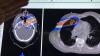
On the workflow side, several vendors are leveraging AI to pull together all of a patients' information, prior exams and reports in one location and to digest the information so it is easier for the radiologist to consume. Often the AI pulls only data and priors that relate to a specific question being asked, based on the imaging protocol used for the exam. One example of this is the Siemens Healthineers AI Clinical Pathway and Siemens AI integrations with PACS to automate measurements and advanced visualization.

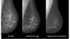
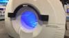
AI is also helping simplify complex tasks and help reduce the reading time on involved exams. One example of this is in 3-D breast tomosythesis with hundreds of images, which is rapidly replacing 2-D mammography, which only produces 4 images. Another example is automated image reconstruction algorithms to significantly reduce manual work. AI also is now being integrated directly into several vendors' imaging systems to speed workflow and improve image quality.

Vendors say AI is here to stay. They explain the future of AI will be automation to help improve image quality, simplify manual processes, improved diagnostic quality, new ways to analyze data, and workflow aids that operate in the background as part of a growing number of software solutions. 

Several vendors at RSNA 2020 noted that AI's biggest impact in the coming years will be its ability to augment and speed the workflow for the small number of radiologists compared to the quickly growing elder patient populations worldwide. There also are applications in rural and developing countries were there are very low numbers of physicians or specialists.


Related AI in Medical Imaging Content:

AI Outperforms Humans in Creating Cancer Treatments, But Do Doctors Trust It?

VIDEO: Artificial Intelligence For MRI Helps Overcome Backlog of Exams Due to COVID

How AI is Helping the Fight Against Breast Cancer

VIDEO: Use of Artificial Intelligence in Nuclear Imaging

3 High-impact AI Market Trends in Radiology at RSNA 2019


Photo Gallery of New Imaging Technologies at RSNA 2019

VIDEO: Editors Choice of the Most Innovative New Radiology Technology at RSNA 2019

Study Reveals New Comprehensive AI Chest X-ray Solution Improves Radiologist Accuracy

VIDEO: Real-world Use of AI to Detect Hemorrhagic Stroke

The Radiology AI Evolution at RSNA 2019


Eliminating Bias from Healthcare AI Critical to Improve Health Equity

VIDEO: FDA Cleared Artificial Intelligence for Immediate Results of Head CT Scans

Building the Future of AI Through Data

WEBINAR: Do More, Perform Better: Delivering Clinical Quality through Advanced Radiology and Artificial Intelligence

Integrating Artificial Intelligence in Treatment Planning


Selecting an AI Marketplace for Radiology: Key Considerations for Healthcare Providers

Artificial Intelligence Improves Accuracy of Breast Ultrasound Diagnoses

Artificial Intelligence Greatly Speeds Radiation Therapy Treatment Planning

WEBINAR: Building the Bridge - How Imaging AI is Delivering Clinical Value Across the Care Continuum

AI in Medical Imaging Market to Reach $1.5B by 2024


VIDEO: AI-Assisted Automatic Ejection Fraction for Point-of-Care Ultrasound

5 Trends in Enterprise Imaging and PACS Systems

VIDEO: Artificial Intelligence to Automate CT Calcium Scoring and Radiomics

Scale AI in Imaging Now for the Post-COVID Era

VIDEO: Integrating Artificial Intelligence Into Radiologists Workflow


Northwestern Medicine Introduces Artificial Intelligence to Improve Ultrasound Imaging

Find more artificial intelligence news and video


Coronavirus (COVID-19) | January 26, 2021

This is an example of a COVID-19 (SARS-CoV-2) positive patient's lung computed tomography (CT) scan. The video scrolls through the image slices of the scan and shows the typical white, ground glass opacities (GGO) caused by COVID pneumonia. The pneumonia typically appears along the walls of each lobe of the lung, especially the chest wall and the lower portions of the lungs. This scan is from a Canon Aquilion Prime SP CT scanner and used Advanced intelligent Clear-IQ Engine (AiCE), an artificial intelligence-driven image reconstruction software to improve image quality of lower-dose scans. This was shown by Canon Medical as an exmaple of CT image quality for the virus at the 2020 Radiological Society of North American (RSNA) meeting. 

Read more about this system and its launch in 2020 to address COVID, Canon Medical Launches CT Solution for Patients with Viral Infectious Diseases.

VIDEO: How to Image COVID-19 and Radiological Presentations of the Virus interview with Margarita Revzin, M.D., associate professor of radiology and biomedical imaging, Yale School of Medicine.

Find more radiology clinical images of coronavirus in this photo gallery.

Find more radiology related COVID news and video

Artificial Intelligence | December 02, 2020

Kirti Magudia, M.D., Ph.D., an abdominal imaging and ultrasound fellow at the University of California San Francisco, explains how an automated deep learning analysis of abdominal computed tomography (CT) images can produce a more precise measurement of body composition and better predicts major cardiovascular events, such as heart attack and stroke, better than overall weight or body mass index (BMI). This was according to a study she presented at the 2020 Radiological Society of North America (RSNA) virtual meeting.

Unlike BMI, which is based on height and weight, a single axial CT slice of the abdomen visualizes the volume of subcutaneous fat area, visceral fat area and skeletal muscle area. However, manually measuring these individual areas is time intensive and costly. A multidisciplinary team of researchers, including radiologists, a data scientist and biostatistician, developed a fully automated artificial intelligence (AI) method to determine body composition metrics from abdominal CT images.

Statistical analysis demonstrated that visceral fat area was independently associated with future heart attack and stroke. BMI was not associated with heart attack or stroke. 

Read more about this study

Find more RSNA news

Information Technology | December 01, 2020

Treating cancer effectively often includes a combination of patient therapies. In recent years, technology advancements have led to a more efficient and personalized approach to treatment. Andrew Wilson, President of Oncology Informatics at Elekta, discussed the latest software advancements with ITN.

Remote Viewing Systems | November 28, 2020

Konica Minolta’s theme for RSNA 2020 is Depth of Vision. ITN recently talked with David Widmann, President and CEO of Konica Minolta Healthcare Americas, about this focus and their key messages for customers and RSNA attendees.

X-Ray | November 28, 2020

Agfa is looking to transform X-ray with new advancements in volumetric imaging, and with new mobile concepts and implementation of intelligent tools. ITN had a conversation with Georges Espada on Transforming X-ray with Intelligent Tools. 

Enterprise Imaging | November 23, 2020

Fujifilm's next generation secure server-side viewer platform extends across enterprise imaging areas to bring together radiology, mammography and cardiology into a single zero footprint platform. Bill Lacy, vice president of medical informatics for Fujifilm Medical Systems USA recently talked with ITN about their Synapse 7x platform.

Artificial Intelligence | November 11, 2020

Artificial Intelligence (AI) is becoming more common place in radiology practices, and emerging technologies are providing radiologists with sophisticated detection software to aid their reading and provide support for a busy workflow. With the progression of AI technology, vendors must look not only at what AI can do for the radiologist, but how the radiologist and the technician interact with that technology –  the goal should be increasing accuracy while also positively improving workflow. GE Healthcare is working to improve radiology AI workflow in its Centricity Universal Viewer.

Three key opinion leaders offers their views on what is needed to make AI more valauble and accessible to radiologists. These include:

   • Amy Patel, M.D., breast radiologist, medical director, Liberty Hospital Women's Imaging, assistant professor of radiology, University of Missouri-Kansas City.

   • Prof. Dr. Thomas Frauenfelder, M.D., vice chairman and professor of thoracic radiology, Institute for Diagnostic and Interventional Radiology, University of Zurich.

   • Randy Hicks, M.D., chief executive officer, Regional Medical Imaging.


Learn more about the Centricity Universal Viewer in the VIDEO: How GE Healthcare’s Zero Footprint Remote Image Viewer Supports Clinical Care


Artificial Intelligence | October 26, 2020

GE Healthcare is highlighting artificial intelligence (AI) automation features on its Voluson Swift ultrasound platform at the 2020 Radiological Society of North America (RSNA) virtual meeting. Features of this system include semi-automated contouring, auto identification of fetal anatomy and positioning on imaging, 

The new SonoLyst AI software can auto recognize 20 standard fetal views in the second trimester protocol. The goal is to speed exam times and make the exams more accurate, even for less experienced sonographers. The AI can tell users what any image is when they freeze the frame. This can be used to help cue up measurements and appropriate annotations. The AI also can tell th user if all the required anatomical structures are in an image needed for the exam protocols.
 

Find more RSNA news and video

Subscribe Now