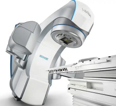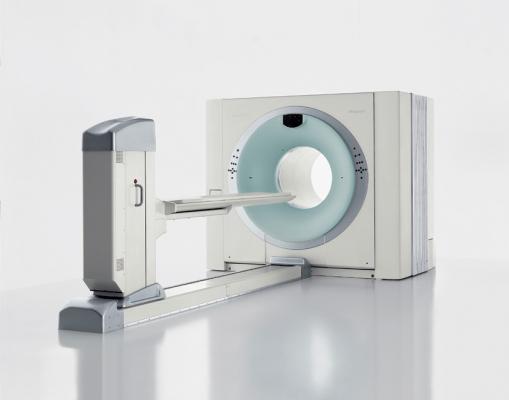
Image courtesy of Siemens Healthcare
November 3, 2015 — The University of Tennessee Medical Center announced it has implemented new software aimed at significantly improving positron emission tomography/computed tomography (PET/CT) image quality and radiation oncology services. It is first time in the world this software is being utilized.
Assistant Professor and Director of UT Medical Center’s Molecular Imaging and Translational Research Program, Dustin Osborne, M.D., explains patients can now benefit from the newly released VG60 software update to the Siemens Biograph 64 mCT Flow PET/CT units utilized inside the medical center.
“UT Medical Center was chosen to pioneer this PET/CT software because of our unique workflows that blend standard oncological imaging with radiation therapy,” said Osborne. “This software contains a number of new features that result in improved radiation therapy services. Plus, combining standard PET/CT and radiation oncology workflows provides more convenience for patients as they can have multiple procedures performed during one appointment.”
According to Osborne, VG60 utilizes features that improve the image quality of PET/CT scans, which have previously been distorted by software limitations and common human error.
“A PET/CT scan is sensitive to any movement or abnormality in the human body which can blur the images,” said Osborne. “The simple respiratory motion of a patient during a scan can have a negative impact on accurate cancer diagnosis and therapy monitoring.”
Osborne explains that this software is significant in the benefits it provides as it has the ability to remove metal artifacts such as implants or dental work from the diagnostic CT images. Patients, physicians and clinicians viewing the scans will collectively benefit from improved large field of view imaging as well as respiratory motion correction that can be added seamlessly to any standard whole-body image study.
“This software solves a number of challenges for using PET/CT in radiation oncology while also vastly improving diagnostic image quality,” said Osborne. “Most importantly, it helps in the expedited diagnosis of our patients. The fact that UT Medical Center is the first in the world to utilize the software is a testament to our physicians and the thousands of patients that annually employ our radiological services.”
For more information: www.utmedicalcenter.org

 April 10, 2024