September 1, 2015 — University of Maryland Medicine and its Center for Metabolic Imaging and Image-Guided Therapeutics (CMIT) has begun to use magnetic resonance imaging (MRI)-guided focused ultrasound on a deep structure within the brain related to Parkinson’s disease.
In the first clinical trial of its kind, researchers from the departments of diagnostic radiology & nuclear medicine, neurosurgery and neurology at CMIT are using MRI to guide ultrasound waves through the intact skin and skull to the globus pallidus. The University of Maryland is one of only two sites in the United States to offer this treatment to Parkinson’s patients.
The globus pallidus contributes to the regulation of voluntary movements and is targeted with medications and, in advanced cases, deep brain stimulation using implanted micro-electrodes to treat motor symptoms of tremor, rigidity and dyskinesia in patients with Parkinson’s. Dyskinesia (abnormal, distorted movement) is a common side effect of the medication levodopa that can affect quality of life for patients with Parkinson’s.
“In collaboration with my colleagues, we are excited to offer our patients a new, non-invasive therapy to control their Parkinson’s symptoms,” said principal investigator Howard M. Eisenberg, M.D., the Raymond K. Thompson Chair of Neurosurgery. “The neurology community has made significant strides in helping patients with Parkinson’s over the years; utilization of MRI-guided focused ultrasound could help limit the life-altering side effects like dyskinesia to make the disease more manageable and less debilitating.”
“For years, our medical techniques have centered around anatomic imaging of the body and open surgical techniques to repair structural problems,” said Graeme Woodworth, M.D., associate professor of neurosurgery and director of the neurosurgery department’s Translational Research Laboratory. “CMIT is set to move this paradigm toward imaging body function and modifying alterations using non-invasive, image-guided focused ultrasound technology. We are very excited that these new technologies, available at the University of Maryland, are on course to revolutionize medical diagnosis and treatment,” he said.
According to Woodworth, examples of this work leverage altered metabolic pathways to visualize disease processes and treatment responses, providing new information regarding the course of a patient's condition. Using advanced focused ultrasound technology, surgeons can now apply microscopic sound waves to precisely target diseased regions deep within the body without incisions or radiation.
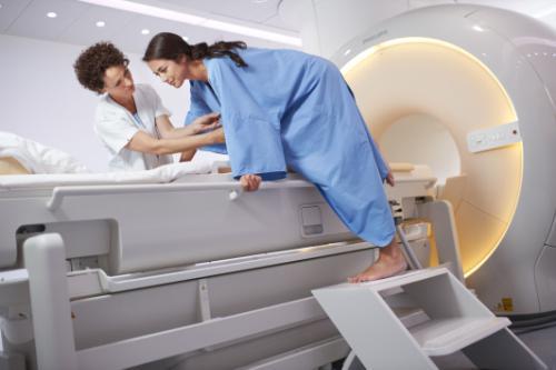
Functional imaging and non-invasive ultrasound procedures are done on an outpatient basis in the CMIT MRI suite. During the Parkinson’s procedure, patients lie in an MRI scanner with a head-immobilizing frame fitted with a transducer helmet. Ultrasonic energy is targeted through the skull to the globus pallidus of the brain, and images acquired during the procedure give physicians a real-time map of the area being treated.
“We’re raising the temperature in a very restricted area of the brain to destroy tissue,” Dr. Eisenberg said. “The ultrasound waves create a heat lesion that we can monitor through MRI.”
The entire procedure lasts two to four hours, and patients are awake and able to interact with the treatment team. This allows the physicians to monitor the immediate effects of treatment and make adjustments if necessary.
“Treatment-related side effects such as dyskinesia are the main reason my patients undergo surgery,” added Paul S. Fishman, M.D., Ph.D., professor of neurology and sub-investigator on the clinical trial. “Focused ultrasound could offer these patients an alternative to surgery.”
The clinical study builds on experience gained during a pilot trial that investigated focused ultrasound for patients with essential tremor. The University of Maryland Medical Center (UMMC) was one of eight sites that participated in the pivotal Phase III trial to support a submission to the U.S. Food and Drug Administration (FDA) for regulatory approval.
Researchers from the University of Virginia Health System reported in the New England Journal of Medicine in 2013 that 15 patients with essential tremor who received focused ultrasound saw “significant improvement” in their dominant hand tremor. Patients treated in the initial phase of the study at the University of Maryland experienced similar results.
The Michael J. Fox Foundation for Parkinson’s Research and the Focused Ultrasound Foundation are funding the Parkinson’s study. It is being conducted using the ExAblate Neuro system developed by InsighTec.
For more information: www.medschool.umaryland.edu

 April 23, 2024


