February 2, 2016 — Researchers are using a mathematical tool to help determine which concussion patients will go on to suffer migraine headaches, according to a new study published online in the journal Radiology.
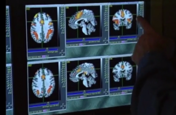
Post-traumatic migraine headaches are common in people who suffer concussions. In recent years, researchers have been using diffusion tensor imaging, a type of magnetic resonance imaging (MRI), to assess concussion-related damage to the brain’s signal-transmitting white matter and look for associations with symptoms like headaches. MRI can be used to create a frequency distribution graph of the whole brain called a histogram, and from that a mean fractional anistropy (FA), a measure of how easily water moves through the brain, can be derived to assess white matter injury. However, mean FA has shortcomings.
“Mean FA represents an average,” said study author Lea M. Alhilali, M.D., from the University of Pittsburgh Medical Center in Pittsburgh. “If someone has a higher FA to begin with and they lose white matter integrity from trauma, they still might average out to have a normal mean FA.”
Instead, the researchers analyzed the MRI results using information theory, a branch of mathematics based on mathematical laws surrounding the behavior of data as it is retrieved, transferred or stored. Shannon entropy, an information theory model that looks at areas of entropy, or disorder, in a complex system like the brain, has advantages over mean FA in the analysis of brain histograms, according to Alhilali.
“A healthy brain has high entropy, but people with injuries to the white matter from trauma may lose some of that complexity and have less entropy,” she explained.
In the first study of its kind, Alhilali and colleagues assessed the performance of Shannon entropy as a diagnostic tool in concussion patients with and without post-traumatic migraines.
They obtained FA maps and neurocognitive testing results in 74 concussion patients, including 57 with post-traumatic migraines and 17 without. FA maps were obtained in 22 healthy controls and 20 control patients with migraine headaches for comparison. Mean FA and Shannon entropy were extracted from total brain FA histograms and compared between concussion patients and controls and between those with and without post-traumatic migraine.
Shannon entropy analysis of FA histograms performed better than mean FA as a diagnostic test to differentiate between concussion patients and controls and also performed better in determining which concussion patients developed post-traumatic migraines. The concussion patients had significantly lower Shannon entropy compared to controls, and those with post-traumatic migraines had significantly lower Shannon entropy than other concussion patients. Patients with Shannon entropy below 0.750 were approximately 16 times more likely to have experienced concussion and three times more likely to develop post-traumatic migraines.
Shannon entropy inversely correlated with time to recovery, meaning that people with lower entropy took longer to recover.
The results suggest that Shannon entropy may provide a convenient, reproducible biomarker that can be calculated in automated fashion to help triage patients after initial injury and predict which ones will go on to get more severe symptoms.
“This approach requires just one histogram for the entire brain,” Alhilali said. “If it continues to show promise, then it could be added to the regular brain MRI as part of the study.”
Additional research is needed to study other potential applications of Shannon entropy, Alhilali said, such as predicting future cognitive performance in concussion patients.
For more information: www.radiologyinfo.org

 April 23, 2024


