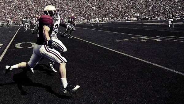
April 26, 2016 — Repeated head impacts to high school football players cause measurable changes in their brains, even when no concussion occurs, according to new research. The study was conducted by University of Texas Southwestern Medical Center’s Peter O’Donnell Jr. Brain Institute and Wake Forest University School of Medicine.
Researchers gathered data from high school varsity players who donned specially outfitted helmets that recorded data on each head impact during practice and regular games. They then used experimental techniques to measure changes in cellular microstructure in the brains of the players before, during and after the season.
“Our findings add to a growing body of literature demonstrating that a single season of contact sports can result in brain changes regardless of clinical findings or concussion diagnosis,” said senior author Joseph Maldjian, M.D., chief of the Neuroradiology Division and director of the Advanced Neuroscience Imaging Research Lab, part of the Peter O’Donnell Jr. Brain Institute at UT Southwestern.
In the study, appearing in the Journal of Neurotrauma, a team of investigators at UT Southwestern, Wake Forest University Medical Center and Children’s National Medical Center evaluated about two dozen players over the course of a single football season. The group of players was not large enough to draw conclusions about the differences in impacts between positions, researchers said, and additional studies will be needed to determine what the deviations mean clinically for individuals.
“Studies like this are important to understand how and where long-term damage might be occurring, so that we can then take the necessary steps to prevent it,” said first author Elizabeth Davenport, M.D., a postdoctoral researcher in the Department of Radiology and the Advanced Imaging Research Center at UT Southwestern.
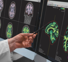
During the pre-season each player had a magnetic resonance imaging (MRI) scan and participated in cognitive testing, which included memory and reaction time tests. During the season they wore sensors in their helmets that detected each impact they received. Post-season, each player had another MRI scan and another round of cognitive tests.
Researchers then used diffusional kurtosis imaging (DKI), which measures water diffusion in biological cells, to identify changes in neural tissues. DKI analysis has been used to detect changes in neural tissues to study brain development, as well as brain injury and disease including autism spectrum disorders, attention deficit/hyperactivity disorder (ADHD), Alzheimer’s disease, traumatic brain injury (TBI), stroke, schizophrenia and mild cognitive impairment.
DKI also allowed the researchers to measure white matter abnormalities. White matter consists of fibers that connect brain cells and can speed or slow signaling between nerve cells. In order for the brain to reorganize connections, white matter must be intact and the degree of white matter damage may be one factor that limits the ability of the brain to reorganize connections following TBI.
“Work of this type, combining biomechanics, imaging and cognitive evaluation is critical to improving our understanding of the effects of subconcussive impacts on the developing brain,” said Maldjian, professor of radiology and the Advanced Imaging Research Center at UT Southwestern. “Using this information, we hope to help keep millions of youth and adolescents safe when engaged in sports activities.”
Football has the highest concussion rate of any competitive contact sport, and there is growing concern – reflected in the recent decrease in participation in the Pop Warner youth football program – among parents, coaches and physicians of youth athletes about the effects of subconcussive head impacts, those not directly resulting in a concussion diagnosis, researchers noted. Previous research has focused primarily on college football players, but recent studies have shown impact distributions for youth and high school players to be similar to those seen at the college level, with differences primarily in the highest impact magnitudes and total number of impacts, the researchers noted.
The findings contribute to a growing body of knowledge and study about concussions and other types of brain injury by researchers with the Peter O’Donnell Jr. Brain Institute. Among them:
- In the first study of its kind, former National Football League (NFL) players who lost consciousness due to concussion during their playing days showed key differences in brain structure later in life. The hippocampus, a part of the brain involved in memory, was found to be smaller in 28 former NFL players as compared with a control group of men of similar age and education;
- A study examining the neuropsychological status of former NFL players found that cognitive deficits and depression are more common among retired players than in the general population;
- CON-TEX includes one of the nation’s first registries of concussion patients ages 5 and older to capture comprehensive, longitudinal data on sports-related concussion and mild traumatic brain injury patients; and
- The Texas Institute for Brain Injury and Repair (TIBIR), a state-funded initiative to promote innovative research and education in traumatic brain injury, includes a comprehensive Concussion Network that delivers expert brain injury education to coaches, school nurses, athletic trainers and parents about the risks of sports-related injuries.
In this study, other institutions involved included Wake Forest University, the Children’s National Medical Center at George Washington University School of Medicine and Medical University of South Carolina.
Support for the study included the National Institutes of Health, Childress Institute for Pediatric Trauma at Wake Forest Baptist Medical Center and a National Science Foundation Graduate Research Fellowship grant.
For more information: www.liebertpub.com/overview/journal-of-neurotrauma


 April 17, 2024


