November 21, 2016 — Parents may soon be able to watch their unborn babies grow in realistic 3-D immersive visualizations, thanks to new technology that transforms magnetic rasonance imaging (MRI) and ultrasound data into a 3-D virtual reality model of a fetus, according to research being presented next week at the 2016 meeting of the Radiological Society of North America (RSNA).
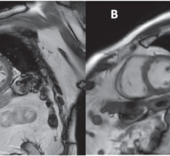
MRI provides high-resolution fetal and placental imaging with excellent contrast. It is generally used in fetal evaluation when ultrasound cannot provide sufficiently high-quality images.
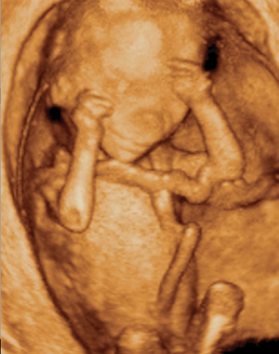
Researchers in Brazil created virtual reality 3-D models based on fetal MRI results. Sequentially-mounted MRI slices are used to begin construction of the model. A segmentation process follows in which the physician selects the body parts to be reconstructed in 3-D. Once an accurate 3-D model is created — including the womb, umbilical cord, placenta and fetus — the virtual reality device can be programmed to incorporate the model.
"The 3-D fetal models combined with virtual reality immersive technologies may improve our understanding of fetal anatomical characteristics and can be used for educational purposes and as a method for parents to visualize their unborn baby," said study co-author Heron Werner Jr., M.D., Ph.D., from the Clínica de Diagnóstico por Imagem, in Rio de Janeiro, Brazil.
The virtual reality fetal 3-D models are remarkably similar to the postnatal appearance of the newborn baby. They recreate the entire internal structure of the fetus, including a detailed view of the respiratory tract, which can aid doctors in assessing abnormalities.
For the virtual reality device, Werner and colleagues used the latest-generation Oculus Rift 2 headset. Oculus Rift 2 places the user in an immersive environment, complete with heartbeat sounds derived from the ultrasound of the fetus. Users can study the 3-D fetal anatomy simply by moving their head.
"The experience with the Oculus Rift has been wonderful," Werner said. "It provides fetal images that are sharper and clearer than ultrasound and MR images viewed on a traditional display."
The technology has numerous potential applications, including assessment of fetal airway patency. Airway patency, or the state of airways being open and unblocked, is an important issue for a developing fetus. For example, if ultrasound showed an abnormal mass near the fetal airway, physicians could use the 3-D images and the headset to assess the entire length of the airway and make better informed decisions about delivery.
The technology also can help coordinate care with multidisciplinary teams and provide better visual information to parents to help them understand malformations and treatment decisions.
"The physicians can have access to an immersive experience on the clinical case that they are working on, having the whole internal structure of the fetus in 3-D in order to better visualize and share the morphological information," Werner said. "We believe that these images will help facilitate a multidisciplinary discussion about some pathologies in addition to bringing a new experience for parents when following the development of their unborn child."
The researchers have used the technique on patients at a clinic in Rio de Janeiro, including cases where the fetus had evidence of an abnormality that required postnatal surgery. They hope to use the technology more broadly over the next year.
Co-authors on the study are Bianca Guedes Ribeiro, M.D., Jorge Lopes, Ph.D., Gerson Ribeiro, Pedro Daltro, M.D., Tatiana M. Fazecas, M.D., Renata A. Nogueira, M.D., and Leise Rodrigues, M.D.
For more information: RadiologyInfo.org

 April 17, 2024