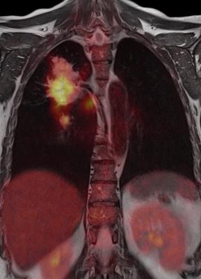
August 2, 2016 — A cutting-edge method of extracting big data from positron emission tomography (PET) images can provide additional information to quantify lung tumors caused by a genetic mutation. This information could help guide the most effective treatment, suggest findings of a study of nearly 350 patients being presented at the 58th Annual Meeting of the American Association of Physicists in Medicine (AAPM), July 31-Aug. 4 in Washington, D.C.
Advances in genomics – the analysis of an organism’s genetic information – have shown that non-small cell lung cancer (NSCLC), the most common type of lung cancer, often is caused by mutations in specific genes. These mutations include epidermal growth factor receptor (EGFR) and Kristen rat sarcoma viral (KRAS). For example, 15 percent of patients have EGFR mutations and often benefit from tyrosine kinase inhibitor (TKI) therapies. Therefore, identification of these mutations is crucial for selecting the most effective treatment for these patients.
PET imaging often is used to assess tumor glucose metabolism and an essential tool for lung cancer management. Studies have shown that some of the biological and genetic variations within tumors potentially can be captured in PET images.
Using big data radiomics — extracting comprehensive information from PET images — researchers evaluated the associations between radiomic features of tumors and EGFR and KRAS mutations in about 350 NSCLC lung cancer patients. The mutations were confirmed by molecular testing based on biopsies of tumor tissues, the standard of care for mutation identification. Researchers found that radiomic features describing different aspects of the tumor, such as its shape and textures, appear to be associated with EGFR mutations. Their results suggest that different metabolic imaging patterns (or imaging phenotypes) that are quantified by radiomic features may be caused by EGFR mutations.
“Our long-term goal would be to use PET or another imaging technique to develop non-invasive imaging biomarkers that complement molecular tests,” said Hugo Aerts, Ph.D., director of the Computational Imaging and Bioinformatics Laboratory at Dana Farber Cancer Institute, Brigham and Women’s Hospital and Harvard Medical School in Boston.
“Medical images are regularly acquired for every cancer patient that comes into our clinic for treatment, which is the case at many other cancer centers as well,” said Stephen Yip, Ph.D., an instructor at Harvard Medical School, Dana Farber Cancer Institute and Brigham and Women’s Hospital. “This early research suggests that standard-of-care PET imaging may help guide doctors in identifying patients with EGFR mutations, potentially providing valuable information for personalized lung cancer therapy.”
“More research needs to be done to further understand the complementary role of PET imaging to molecular testing,” Aerts said.
The researchers are now assessing combining PET-based radiomic features with features derived from other imaging tests, such as computed tomography (CT) or magnetic resonance imaging (MRI), to improve the accuracy of genetic mutation identification in lung and brain cancers.
For more information: www.aapm.org


 April 17, 2024
