February 3, 2016 — A new way of using magnetic resonance imaging (MRI) scanners to look for evidence of multiple sclerosis in the brain has been successfully tested by researchers at The University of Nottingham and Nottingham University Hospitals NHS Trust.
Multiple sclerosis (MS) is a neurological condition which affects around 100,000 people in the United Kingdom. It is notoriously difficult to diagnose as it has many symptoms but not all sufferers experience all of them and the disease can progress at different rates. MRI scans have been used as a diagnostic tool to detect white matter lesions in the brain but these are not always an indicator of the disease.
Now a research team at Nottingham has found a way to use clinical MRI to distinguish between MS lesions and other brain white spots which are found in MS. The study is published in the Multiple Sclerosis Journal.
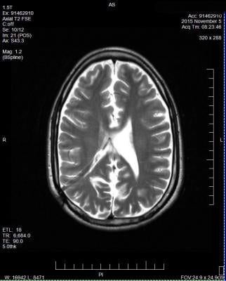
They have used a clinical MRI scanner of the type all neuroscience centers have to carry out a special type of scan called a T2-weighted imaging process which is able to reveal lesions in the brain's white matter that are centered on a vein — a known indicator of MS.
Leading the work, Nikos Evangelou, M.D., said: "We already knew that large research MRI scanners could detect the proportion of lesions with a vein in the brain's white matter, but these scanners are not clinically available. So we wanted to find out whether a single brain scan in an NHS hospital scanner could also be effective in distinguishing between patients known to have MS and patients known to have non-MS brain lesions. We are excited to reveal that our results show that clinical application of this technique could supplement existing diagnostic methods for MS."
A total of 40 patients were recruited from the neurology outpatients' department of Nottingham University Hospitals NHS Trust. Initially a test cohort of 10 patients with MS and 10 patients with non-MS white brain matter lesions were scanned. Anonymized scans were analyzed blinded to clinical data and simple diagnostic rules were devised. The same rules were applied to a validation cohort of 20 patients (13 with MS and 7 with other lesions) by a blinded observer.
Within the test cohort, all patients with MS had central veins in more than 45 percent of brain lesions, while the rest had central veins visible in less than 45 percent of lesions. Then, by applying the same diagnostic rules to the second cohort, all the remaining patients were correctly categorized into MS or non-MS, by the blinded observer, taking less than two minutes per scan.
The new study is significant because currently among patients referred to MS treatment centers with suspected MS, fewer than 50 percent are found to have it. This shows that diagnosing MS in a significant minority of cases can be challenging.
The Nottingham University team has now started a new study examining patients with real uncertainty about the diagnosis and aim to extend the study in other U.K. towns so more patients can participate. It is possible that in less than two years they will know if this new test is accurate as it appears to be.
The Nottingham team has already presented their data in the United States and a similar U.S.-based study is planned based on the Nottingham results.
For more information: www.msj.sagepub.com

 April 24, 2024


