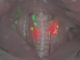
Intraoperative image of thyroid (red) and parathyroids (green) in rats four hours after intravenous injection of targeted contrast agents. Image captured using the FLARE system. Image courtesy of Macmillan Publishers Ltd.
July 29, 2015 — Researchers funded by the National Institute of Biomedical Imaging and Bioengineering (NIBIB) have developed two near-infrared (NIR) contrast agents that are efficiently taken up by the thyroid and parathyroid glands following intravenous injection. The researchers tested the contrast agents in rats and pigs and showed that they could be used to help distinguish the thyroid and parathyroid glands from surrounding tissue and from each other. Their findings appeared in the Jan. 5, 2015, issue of Nature Medicine.
The thyroid is a butterfly-shaped gland located in the front of the neck that controls the rate of many activities involved in metabolism. Directly behind the thyroid sit four parathyroid glands, which tightly control calcium levels in the blood; each is approximately the size of a grain of rice.
If the thyroid isn’t working properly, or if a tumor is present, surgery may be needed to remove all or part of the gland. While the thyroid and parathyroids can be located prior to surgery via ultrasound, once a patient is on the operating table surgeons have only their eyes and knowledge of anatomy to help them distinguish these important glands from other tissues in the body. Inability to do so can result in incomplete removal of the thyroid or inadvertent damage to the parathyroids, which can cause chronically low calcium levels.
“Proper identification of the thyroid and parathyroid glands during head and neck surgery is critical for avoiding accidental injury, but presents a significant challenge due to their small size and variations in location from patient to patient,” says Richard Conroy, Ph.D., director of the Division of Applied Science & Technology at NIBIB. “The innovative design and optimization of these contrast agents has the potential to lead to the development of other tissue-specific small molecule contrast agents. It’s a great example of how investing in the development of new imaging technologies can help overcome existing challenges in medicine.”
Upon intravenous injection, the new contrast agents are taken up by the thyroid and parathyroids, where they emit different wavelengths of NIR light. Although this light is invisible to the naked eye, it can be easily seen using a specially-designed NIR camera called FLARE. The FLARE camera shoots a color video of the surgical site and also captures the two NIR wavelengths to create a unique view in which the tissues that have taken up contrast agents glow brightly. In their experiments on rats and pigs, the researchers used FLARE to visualize the thyroid and parathyroid simultaneously.
The development of the contrast agents was led by John V. Frangioni, M.D., Ph.D., professor of medicine at Harvard Medical School, and Hak Soo Choi, Ph.D., associate professor of medicine at Harvard Medical School; chemical synthesis was performed at Georgia State University in the laboratory of Maged Henary, Ph.D.
Frangioni is also CEO of Curadel, a company he started in 2014 with the purpose of developing NIR contrast agents and imaging systems to help surgeons identify glands, blood vessels, nerves, lymph nodes, and tumors during operations.
“We’re giving surgeons the ability to find these structures through blood and through tissue, and in doing so, we’re bringing surgery into the 21st century,” said Frangioni.
The novel thyroid and parathyroid contrast agents differ significantly from most current NIR contrast agents, which are composed of two separate molecules linked together: a molecule that emits a fluorescent signal, called the fluorophore, and an antibody or small molecule that targets the tissue of interest.
“You can think of the fluorophore as a lightbulb,” said Frangioni. “The conventional way to create a targeted fluorophore is to take a targeting ligand, such as an antibody, and hook it up to the lightbulb to create a new hybrid molecule. That’s the way people have been doing it for 50 years.”
But hybrid molecules tend to be large, and large molecules aren’t easily cleared by the body. This is a problem, said Frangioni, because when fluorescent molecules are injected into the blood, only a small fraction are taken up by the tissue of interest; the rest continue to circulate through the blood until filtered either by the kidneys or liver and excreted from the body. If a molecule is too big to be filtered, it will stay in the bloodstream and continue to emit fluorescent signal. This background signal makes it harder for surgeons to see signal being emitted from the structure of interest.
With the new type of contrast agents, the targeting component is incorporated directly into the chemical structure of the fluorophore to reduce the overall size and encourage clearance. “We basically deconstruct the targeting molecule into little pieces and scatter them around the fluorophore so that in three dimensions it’s a very compact molecule,” said Frangioni, who coined the novel arrangement “structure-inherent targeting.”
Frangioni said another advantage of a single molecule as opposed to a larger hybrid molecule is that, in the future, it will be less difficult to manufacture. “It’s a lot easier to make a single molecule instead of having to make the lightbulb and make the targeting molecule and put the two together. For those of us who are developing drugs for commercialization, that’s a big deal. If you can cut the number of steps in half, that’s huge.”
For more information: www.nature.com

 April 18, 2024


